Download
1 / 42
420 likes | 665 Vues
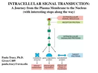
INTRACELLULAR SIGNAL TRANSDUCTION: A Journey from the Plasma Membrane to the Nucleus (with interesting stops along the way). Paula Tracy, Ph.D. Given C409 paula.tracy@uvm.edu. INTRACELLULAR SIGNALING: Signal Transduction.

E N D
INTRACELLULAR SIGNAL TRANSDUCTION: A Journey from the Plasma Membrane to the Nucleus (with interesting stops along the way) Paula Tracy, Ph.D. Given C409 paula.tracy@uvm.edu
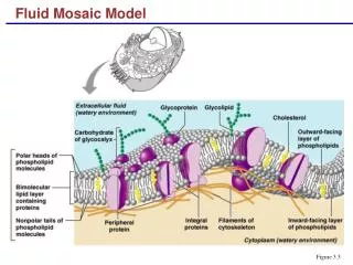
INTRACELLULAR SIGNALING: Signal Transduction Cell membranes, as well as the cell cytoplasm and even the cell nucleus, contain cell-specific receptors for various ligands, which are involved in outside-inside signaling, i.e. signal transduction. Ligands include hormones, growth factors, cytokines, prostaglandins and proteases. Hormones are involved in a variety of metabolic processes that maintain homeostasis e.g. fuel metabolism. Particularly noteworthy in that regard are glucagon, insulin and the catecholamines (epinephrine and norepinephrine). Growth factors are involved in mitogenesis, whereas cytokines play critical roles in the differentiation, proliferation and function of various cell lineages.. Interaction of such ligands with their membrane, cell-specific receptors or intracellular receptors causes conformational changes in the receptor and, in many instances receptor- associated cytoplasmic proteins. Such events result in the initiation of a cascade of important, but as yet incompletely understood, events leading to e.g. enzyme activation, differentiation and/or cell division.
General Schemes of Intercellular Signalling Extracellular signaling molecules released by cells occurs over distances from a few microns - autocrine (c) and paracrine (b) signaling to several meters in endocrine (a) signaling. In some instances, receptor proteins attached to the membrane of one cell interact directly with receptors on an adjacent cell (d). © 2000 by W. H. Freeman and Company. All rights reserved.
FORCES DRIVING SELECTION OF CURRENT MECHANISMS Need for coordinated intercellular communication Need to translate extracellular signals into series of intracellular events, while allowing for specificity Specificity Determinants: 1. Specific receptors on or in the target cells recognize an appropriate ligand. 2. Specific response to receptor occupancy - effector pathways Diversity of intercellular communication is achieved with hundreds of signaling molecules, including... Proteins, small peptides, amino acids, nucleotides, steroids, fatty acid derivatives, and even dissolved gases such as NO and CO
Receptor Classes Intracellular receptors: - signaling molecules include steroid hormones, retinoids, thyroxine, etc - receptor-hormone complex acts a transcription factor to alter transcription of certain genes • Cell surface receptors: • - signaling molecules include • peptide hormones, catechol- • amines, insulin, growth • factors, cytokines, etc • binding, and subsequent events, • triggers an or in the • cytosolic concentration of • a second messenger; or the • activated,bound receptor acts • as a scaffold to recruit and • activate other intracellular • proteins © 2000 by W. H. Freeman and Company. All rights reserved.
ADVANTAGES 1. Each cell is programmed to respond to specific combinations of signaling molecules. 2. Different cells can respond differently to the same chemical signal. Molecular Biology of the Cell, 2002
HORMONES - First class of signaling molecules defined Secreted from endocrine cells - specialized signaling cells that control the behavior of an organism as a whole: 1. Differ from other intracellular mediators 2. Usually stimulate metabolic activities in tissues remote from the secretory organ 3. Active at very low concentrations (pM - M) 4. Response to hormonal signal comes as a direct and rapid result of its secretion 5. Metabolized rapidly so effects are, in most instances, short-lived, leading to rapid adaptations to metabolic changes Categories: 1. Peptides or polypeptides - insulin, glucagon, growth hormone, insulin-like growth factors, vasopressin, prolactin…. 2. Glycoproteins - follicle stimulating hormone (FSH), thyroid stimulating hormone (TSH)… 3. Steroids - glucocorticoids (aldosterone, cortisol), steroids (progesterone, testosterone), retinoic acid… 4. Amino acid derivatives - epinephrine, norepinephrine, thyroxine, triidothyronine
AGONISTS vs. ANTAGONISTS Agonistmimics a hormone in binding productively to a receptor Antagonistmimics a hormone stereochemically, but binds to the receptor non-productively,inhibiting the action of the natural hormone Hormone binds 2 receptor in lung bronchial relaxation Agonist e.g. important therapy in asthma Antagonist control heart beat binds 2 receptor in heart muscle increased heart rate
RECEPTOR CHARACTERISTICS 1. Participates in transduction of the signal from the external messenger to some component of the metabolic machinery 2. Has at least one additional functional site which is altered by ligand binding (allosteric site) 3. Ligand binding to receptors is saturable, resembling Michaelis-Menten kinetics © 2000 by W. H. Freeman and Company. All rights reserved. 4. Ligand-receptor interaction characterized by tight binding (Kd = pM - M)
How to calculate a Kd and number of receptors from direct binding data Derivation of a Scatchard Plot
Molecular Biology of the Cell, 2002 Simple Intracellular Signaling Induced by an Extracellular Signaling Molecule A signaling molecule activates its receptor activation of an intracellular signaling pathway, i.e. a series of signaling proteins, which may interact with a target protein to change the behavior of the cell.
Glucagon Epinephrine Thrombin Insulin Growth factors Molecular Biology of the Cell, 2002 THREE LARGEST CLASSES OF CELL SURFACE RECEPTORS
Intracellular Signaling Mediated by G protein-linked Membrane Receptors e.g. Glucagon, Epinephrine and Thrombin as signaling molecules 1. Activates a chain of events alterations in concentrations of signaling molecules; elaborate sets of interacting molecules that can relay signals from cell surface to the nucleus 2. Components: Receptor; Transducer (G protein): Effector (membrane-bound enzyme); Second messenger (e.g. cAMP); Response (cascade of highly-regulated protein phosphorylations, etc) RECEPTOR: typically a seven transmembrane domain, integral membrane protein Ligand binding domain (extracellular site) -adrenergic receptor All G protein-coupled receptors resemble the -adrenergic receptor in their amino acid sequence and membrane topography. Gs binding site (intracellular site)
Signal Transduction Pathway Activation of Adenylate Cyclase by Gs Receptor: 7 TM domain integral membrane protein Transducer: Gs Effector: adenylate cyclase Second messenger: cAMP
Cycle of G protein dissociation/association: Activation of G proteins involves GTP displacement of GDP bound to the subunit and dissociation of the complex from the subunits, an exchange facilitated by a GEF protein(guanine nucleotide exchange factor) specific for Gs. G protein activation The active subunit binds to and activates/ stimulates membrane-associated adenylate cyclase which catalyzes the conversion of ATP to cyclic AMP (cAMP). The GTPase activity associated with the subunit slowly hydrolyzes the bound GTP to GDP which is facilitated by a GAP (GTPase activating protein) specific for Gs. Adenylate cyclase activation GTP hydrolysis The subunit with GDP bound reassociates with the subunits, and membrane associated adenylate cyclase returns to its basal activity level
Characteristics of G proteins 1. G protein is an trimeric protein which binds guanine nucleotides. 2. They function to couple integral membrane receptors to target membrane-bound enzymes. 3. They can be considered molecular switches wherein… GDP(inactive) GTP(active) + 4.The dissociated subunit expresses GTPase activity. 5. GTPS blocks GTPase activity of GTP.
i with GDP bound Region that changes conformation when GTP is hydrolyzed
Signal Transduction Pathway Activation of Adenylate Cyclase by Gs Receptor: 7 TM domain integral membrane protein Transducer: Gs Effector: adenylate cyclase Second messenger: cAMP Response: PKA regulated protein phosphorylations
Molecular Biology of the Cell, 2002 Activation of cAMP-dependent protein kinase (PKA) cAMP binds to the PKA regulatory subunits conformational changes, which causes their dissociation from the catalytic subunits kinase activation. Release of the catalytic subunits requires the binding of more than two cyclic AMP molecules greatly sharpening the response of the kinase to changes in [cAMP]. PKA is a Ser/Thr kinase with discrete substrate specificity, thus facilitating a cascade of highly regulated protein phosphorylations.
Signal Transduction Pathway Activation of Adenylate Cyclase by Gs Down stream effects: 1. Phosphorylation of regulatory enzymes of metabolic pathways. 2. Increase transcription of certain proteins via phosphorylation of CREB leading to protein syn- thesis of target proteins.
Activated PKA enters the nucleus and phosphorylates CREB (cAMP Response Element Binding protein). Once phosphorylated, CREB recruits the coactivator CBP (CREB Binding Protein). This complex binds to the CREB-binding element to stimulate gene transcription. Molecular Biology of the Cell, 2002 How Increased Intracellular cAMP Leads to Increased Gene Transcription.
Signal Transduction Pathway Activation of Adenylate Cyclase by Gs Early off signals: 1. Gs-associated GTPase activity GTP hydrolysis to GDP leading to re- association of GsGDP and dissociation of the hormone/receptor complex Down stream off signals: 1. cAMP hydrolyzed by phosphodiesterase 2. Ser/Thr phosphatases dephosphorylate the phosphorylated target proteins. **
Desensitization or Endocytosis of GPCR’s Effected by Phosphorylation 1. The ligand activated receptor can be phosphorylated on select Ser/Thr residues by GRK (e.g. BARK - adrenergic receptor kinase). These phosphorylated residues provide a docking site for arrestin resulting in inactivation/desensitization. 2. In some instances, arrestin binding targets the receptor for clathrin-dependent endocytosis. 3. In addition, if the occupied GPCR leads to cAMP, the receptor can also be phosphorylated by PKA leading to its inactivation/densensitization.
Gsvs. Gi Regulation of Adenylate Cyclase Activity Gsstimulates adenylate cyclase Giinhibits adenylate cyclase e.g. epinephrine can increase or decrease intracellular cAMP concentrations, depending upon the receptor to which it binds adrenergic receptors couple to Gs, whereas 2adrenergic receptors couple to Gi
Vasopressin binding to its GPCR activates Gs cAMP and activation of PKA. PKA phosphorylates various proteins the ultimate aggregation of microtubular subunits, which insert as water channels in the luminal plasma membrane to increase the reabsorption of water by free diffusion. Figure 21.38 – Devlin, Textbook of Biochemistry
Images of cAMP Transients in Cultured Aplysia Sensory Neurons. The cell was loaded with a fluorophore that would allow for the quantification of cAMP concentrations within the cell. A: Free cAMP in the resting cell is < 5 X 10⁻⁸ M. B: Stimulation with serotonin, activates adenylate cyclase increasing cytoplasmic cAMP to ~ 1 X 10⁻⁶ M (red), especially within fine processes with a high surface to volume ratio. Thurs, within 20 sec of stimulation, the intracellular [cAMP] increased ~ 20-fold.
Inhibition of Gs and Gi by Bacterial Toxins Cholera toxin effects on Gs: ADP ribosylation of an Arg residue in the s subunit of Gs inhibition of associated GTPase activity Pertussis toxin effects on Gi: ADP ribosylation of a Cys residue in the i subunit of Gi an inability to inhibit adenylate cyclase activity. Thus, both toxins cause increased intracellular cAMP concentrations! © 2000 by W. H. Freeman and Company. All rights reserved.
Gsvs Givs Gq Gs and Gi coupled to adenylate cyclase [cAMP] G q coupled to phospholipase C [Ca2+]
INTRACELLULAR Ca2+ AS A “SECOND” MESSENGER Cells must work very hard to maintain low intracellular [Ca2+] in order for Ca2+ to result in effective intracellular signaling. ** Molecular Biology of the Cell, 2002 Cellular mechanisms that maintain very low intracellular Ca2+ concentrations A:Ca2+ is actively pumped out of the cytosol. B: Ca2+ is pumped into the ER and mitochondria.
Ligand Binding to a GPCR Linked to Gq Phospholipase C Activation Phospholipase C (PLC)-catalyzed hydrolysis of PIP2Formation of2 second messengers ** **
Phospholipase C (PLC)-catalyzed hydrolysis of PIP2Formation of2 second messengers IP3 release of Ca2+ from intracellular stores by binding to an IP3-gated Ca2+ channel in the endoplasmic reticulum. DAG with released Ca2+and membrane-associated phosphatidylserine activates Protein Kinase C (PKC - a Ser/Thr kinase). PKC directly phosphorylates intracellular proteins some of which gene transcription.
Phosphoinositide-activated Second Messenger System Intracellular [Ca2+]
Ca2+ “Second” Messenger System Release from intracellular stores subsequent to PLC-catalyzed hydrolysis of PIP2 1. Must be distinguished from cAMP-induced effects where cAMP activates a variety of Ca2+ channels Ca2+ influx from extracellular milieu (e.g. -adrenergic receptor occupancy in muscle cells Ca2+ influx rate and force of heart beat). 2. Increased intracellular [Ca2+] - Third messenger: Immediate vs. Sustained responses - Ca2+ binds to its ubiquitous intracellular receptor, calmodulin, thereby a) activating Ca2+/calmodulin-dependent kinases (CaM kinases), Ser/Thr kinases b) that have their own sets of substrate proteins, some of which when phosphorylated can or gene transcription.
Increased Intracellular Ca2+: A Third Messenger Calmodulin in a ubiquitous intracellular Ca2+ receptor Ca2+/calmodulin (CaM) complexes activate CaM-dependent kinases (e.g. CaM-kinase II) Molecular Biology of the Cell, 2002
Structure of Ca2+/calmodulin complex based on X-ray diffraction and NMR spectroscopy studies Molecular Biology of the Cell, 2002
Ca2+ Second Messenger System Release from intracellular stores subsequent to PLC-catalyzed hydrolysis of PIP2 1. Must be distinguished from cAMP-induced effects where cAMP activates a variety of Ca2+ channels Ca2+ influx from extracellular milieu (e.g. -adrenergic receptor occupancy in muscle cells Ca2+ influx rate and force of heart beat). 2. Increased intracellular [Ca2+] - Third messenger: Immediate vs. Sustained responses - Ca2+ binds to its ubiquitous intracellular receptor thereby a) activating Ca2+/calmodulin- dependent kinases (CaM kinases), Ser/Thr kinases b) that have their own sets of substrate proteins, some of which when phosphorylated can or gene transcription. - Ca2+ works in concert with DAG and PS to stimulate PKC, a Ser/Thr kinase, that like the CaM kinases has multiple substrates.
Protein Kinase C Activation of Gene Transcription Two Independent Pathways Molecular Biology of the Cell, 2002
Down-regulation of Phosphoinositide-activated Second Messenger System Off-signals: 1. IP3 rapidly dephosphorylated by phosphatases. 2. DAG rapidly hydrolyzed. 3. Ca2+ rapidly pumped out. 4. Ser/Thr phosphatases dephosphorylate PKC and CaM kinase targets.