Download
1 / 27
280 likes | 592 Vues
Bacillus Rod Shaped Bacteria. Staphylococcus grapelike clusters. Diplococcus. Spirillum. Neisseria gonorrhea diplococcus. Streptococcus Chains of coccus. Staphylococcus Note the arrangement of bacteria. What shape of bacteria? What arrangement of bacteria?.

E N D
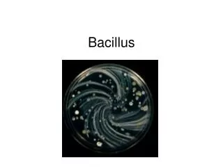
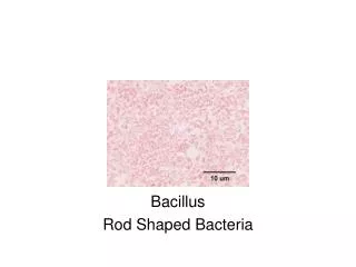
Bacillus Rod Shaped Bacteria
What is meant by a negative stain?Do you observe the capsules around the bacterial cell. Klebsiella pneumonae the causative agent of what disease?
What structure do you observe in the bacterial cells? This is what shape of bacteria?
This is a different type of stain showing what bacterial structure?
What is unusual about the flagella stain process?Note the single flagella at the end called monotrichous.
This flagella pattern is called amphitrichous. It is a tuft of flagella at each end of the bacteria.
Observe the flagella over the entire surface of the bacteria. This pattern is called peritrichous.
This is Rhizopus. In this note the sporangium. The sporangium contains the sporangiospores. The structure which the sporangium is attached is called the sporangiophore.
This is the sexual reproduction of Rhizopus. Note the zygospore in the field of view.
Note the arrangement of the ascus. The spores inside of the ascus are referred to as ascospores. This is a section of Peziza species.
This is Penicillium showing the conidia and the conidiophore. Can you distinguish which one is the conidia and the conidiophore?
This is Aspergillus. Can you identify the conidia? Can you identify the conidiophore?
This is a section of the gill of a basidiomycete. Can you identify the basidiospore, sterigma and basidium.
This is a section through a basidiomycete. Identify the structure indicated in the recipe book on this section.