Download

1 / 29
290 likes | 383 Vues
Understand bradycardia and its management with insights from Pediatric Anesthesiologist, Dr. Alireza Sabzevari. Learn to identify symptoms, differentiate AV blocks, administer atropine, and consider temporary cardiac pacing.

E N D
Bradycardia Dr Alireza Sabzevari,MD Pediatric Anesthesiologist
You arrive at the patient’s room and the nurse informs you that the patient’s HR was 60-70’s during the day but suddenly decreased from 48 to 35bpm. • Current vitals: HR 35bpm, SBP 70/DBP is undetectable RR 16/min, and O2 saturation 93%. • The patient was initially complaining of lightheadedness but now is more lethargic. • You take a look at the EKG that was obtained.
Cornerarstones of managing of bradycardia • Differentiate between signs and symptoms that are caused by the slow rate versus those that are unrelated • Correctly diagnose the presence and type of AV block • Use atropine as the drug intervention of first choice • Deside when to initiate TCP • Deside when to start EP or dopamine to maintain HR and BP • Know when to call for expert consultation about complicated rhythm interpretation, drug , or management decisions
Bradycardia • Bradycardia is defined conservatively as a heart rate below 60 beats per minute, but symptomatic bradycardia generally entails rates below 50 beats per minute. • The 2015 ACLS Guidelines recommend that clinicians not intervene unless the patient exhibits evidence of inadequate tissue perfusion thought to result from the slow heart rate. • Signs & symptoms of inadequate perfusion include hypotension, altered mental status, signs of shock, ongoing ischemic chest pain, & evidence of acute pulmonary edema. Hypoxemia is a common cause of bradycardia; look for signs of labored breathing (eg, increased respiratory rate, retractions, paradoxical abdominal breathing) & low oxygen saturation. Mild symptoms may not warrant treatment.
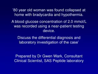
Bradycardias • Many possible causes • Enhanced parasympathetic tone • Increased ICP. • Hypothyroidism • Hypothermia • Hyperkalemia • Hypoglycemia • Drug therapy
Recognition of a symptomatic bradycardia due to AV block is a primary goal. Recognition of the type of AV block is a secondary goal.
Objectives Outline • Normal sinus rhythm • How to recognise an arrhythmia • Bradyarrhythmias • Tachyarrhythmias • Treatment strategy for arrhythmias
How to recognise an arrhythmia How to recognise an arrhythmia • What is the QRS rate? • Are the QRS complexes regular? • Is the QRS broad or narrow? • Are there P waves? • What is the P:QRS relation?
Objectives Outline • Normal sinus rhythm • How to recognise an arrhythmia • Bradyarrhythmias • Tachyarrhythmias • Treatment strategy for arrhythmias
Bradyarrhythmias Bradyarrhythmias • Sinus bradycardia • Sinus arrest (“Sick Sinus Syndrome”) • Junctional bradycardia • Atrioventricular block • (First degree) • Second degree - type I (Wenckebach) / type II • Third degree
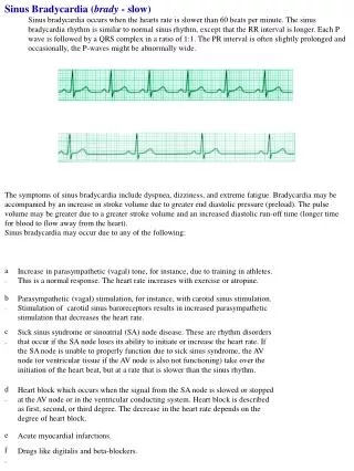
Sinus bradycardia * Rate < 60bpm Regular, narrow QRS P waves present P:QRS is 1:1
Sinus arrest * Rate < 60bpm Irregular, narrow QRS P waves present P:QRS is 1:1 Pause with absence of P wave
Junctional bradycardia * Rate < 60bpm Regular, narrow QRS No P waves
First degree AV block * Rate variable Regular, narrow QRS P waves present P:QRS is 1:1 with PR interval >200ms
Second degree AV block (type I) * * Rate < 60bpm Irregular narrow QRS P:QRS not 1:1 increasing PR interval then dropped beat
Second degree AV block (type II) * * * Rate < 60bpm Irregular narrow QRS P:QRS not 1:1 normal PR interval with intermittent dropped beats
Third degree (complete) AV block * Rate < 60bpm Regular broad QRS No relation between P and QRS
Second degree AV block (type II) * * * Rate < 60bpm Irregular narrow QRS P:QRS not 1:1 normal PR interval with intermittent dropped beats
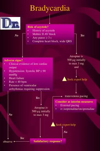
ADULT BRADYCARDIA UNIVERSAL ALGORITHM · Identify and treat reversible causes · Establish vascular access · Obtain 12 lead ECG , when available STABLE UNSTABLE BASE PHYSICIAN ORDER ONLY · Fluid Bolus 500 ml NS · Dopamine 5 - 20 mcg / kg / minute · Sodium Bicarbonate for tricyclic antidepressant OD · Calcium Chloride for suspected hyperkalemia , suspected renal failure or calcium channel blocker OD · Glucagon for beta blocker OD · Atropine Higher doses of for organophosphate OD Adult Bradycardia Be conservative with Atropine. If it does not work, consider other reversible causes rather than giving more Atropine.
Adult Bradycardia Dopamine can be a very effective sympathomimetic for bradycardia not responsive to Atropine. Organophosphate overdoses may require multiple re-boluses of Atropine in order to maintain adequate heart rate.
Adult Bradycardia You arrive on scene at a local restaurant to find a 64 year old male patient sitting in a chair c/o feeling light headed, weak and nauseated. You note the patient is pale and slightly diaphoretic. B/P 100/40, P 50 regular, respirations unlabored at 12. The patient denies any other complaint or medical history. You place the patient on your EKG (Lead II) and find the following rhythm:
Adult Bradycardia After placing the patient on oxygen and establishing an IV, you elect to give the patient 0.5 mg of atropine. Within about one minute, the patient states that the dizziness has subsided, and you note that the patient’s pulse is now 60 and his skin color has improved. Your EKG (Lead II) now looks like this:
Adult Bradycardia You decide since things are going so good, why not do a 12-lead. As you are attaching the 12-lead cables, your patient begins complaining of crushing chest pain and he is now getting very diaphoretic and anxious. You run your 12-lead and now you’re the one getting diaphoretic. Here Is your 12-lead:
Adult Bradycardia THINK TWICE BEFORE GIVING ATROPINE TO BRADYCARDIA PATIENTS WITHOUT 12-LEAD COMPLETED FIRST!!
انواع پيس ميكر الف - داخلی Internal 1-موقت Temporary ب –خارجی (T.C.P) External 2-دائمی Permanent