
SlideServe
Menu
Préférences
© SlideServe · v2026

Ossification = Osteogenesis

loring avatar
13 diapositives · 1.2K Views · May 02, 2012

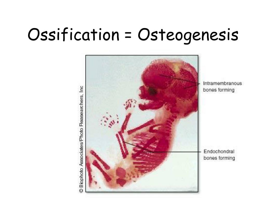
Ossification = Osteogenesis. Ossification = Osteogenesis. Parts of the fetal skeleton form during the first few weeks after conception By the end of the 8 th week, the skeletal pattern is formed : cartilage & connective tissue membranes. There are two classifications of ossification:
Ossification = Osteogenesis - slide 1
01 / 13Ossification = Osteogenesis
1 / 17

Télécharger la présentation

Ossification = Osteogenesis
Une image / un lien ci-dessous est fourni (tel quel) pour télécharger la présentation

Politique de téléchargement : le contenu du site vous est fourni TEL QUEL pour votre information et votre usage personnel ; il ne peut être vendu, concédé sous licence ni partagé sur d’autres sites sans l’accord de son auteur.Le contenu vous est fourni TEL QUEL, pour votre information et votre usage personnel uniquement. Téléchargez la présentation en cliquant sur ce lien. Lors du téléchargement, si vous ne parvenez pas à télécharger une présentation, l’éditeur a peut-être supprimé le fichier de son serveur.Pendant le téléchargement, si vous ne parvenez pas à obtenir une présentation, le fichier a peut-être été supprimé par l’éditeur.

F I N

Plus de contenu associé

Transcription de la présentation
  1. Ossification = Osteogenesis

  2. Ossification = Osteogenesis • Parts of the fetal skeleton form during the first few weeks after conception • By the end of the 8th week, the skeletal pattern is formed : cartilage & connective tissue membranes

  3. There are two classifications of ossification: Intramembranous: Endochondral: *Replacement of *Replacement of sheetlike connective hyaline cartilage tissue with bony tissue with bony tissue *Primarily in flat bones *Most bones are of skull & some irregular formed this way bones

  4. Intramembranous Ossification Osteoblasts migrate to the membrane and deposit bony matrix around themselves. Once surrounded by matrix, they are called osteocytes.

  5. Endochondral Ossification • Hyaline cartilage ‘bones’ are infiltrated with blood vessels & osteoblasts. • Formation of periosteum • ‘Collar’ of compact bone around diaphysis

  6. Endochondral Ossification • At the same time, cartilage in center of diaphysis begins to disintegrate • Replaced with spongy bone * Primary Ossification Center*

  7. Ossification continues from the primary ossification center towards the ends of the bones. After spongy bone is formed in diaphysis, osteoclasts break it down to open up medullary cavity The cartilage in the epiphysis continues to grow, so developing bone increases in length After birth, secondary ossification centers form in epiphyses – ossification is similar, except spongy bone is not broken down. Once ossification is complete – the cartilage is all replaced except at the surface of the epiphysis [articular cartilage] and between the diaphysis and epiphysis[epiphyseal plate]

  8. Secondary Growth Center Epiphyseal Plate  Line

  9. Epiphyseal Growth

  10. Appositional Growth Although bone length growth stops in the ’20s, bones can continue to increase in thickness or diameter throughout life in response to stress from increased muscle activity or weight. Osteoblasts in the periosteum form compact bone around the external bone surface Osteoclasts in the endosteum break down bone around the medullary cavity