
Anatomy of Gluteal Region and Hip Bone
E N D
Presentation Transcript
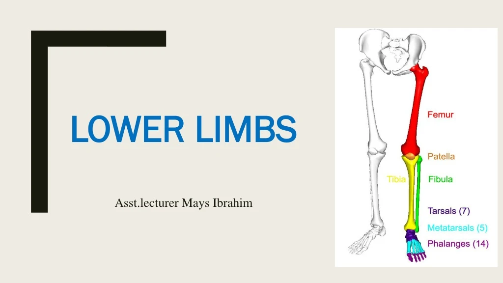
Lower limbs Asst.lecturer Mays Ibrahim
Gluteal region • The buttock or gluteal region is formed mainly by the gluteus maximus muscle. The outline of the superior border of the buttock is formed by the iliac crests.
Surface Features of The Buttock • Gluteus maximus muscle forms the major portion of the prominence of the buttocks. The sciatic nerve is deep to this muscle. • Gluteus medius muscle, superior and lateral to the gluteus maximus muscle. • Gluteal intramuscular injection, another common site for an intramuscular injection is the gluteus medius muscle. In order to give this injection, the buttock is divided into quadrants and the upper outer quadrant is used as an injection site. The iliac crest serves as the landmark for this quadrant. This site is chosen because the gluteus medius muscle in this area is quite thick, and there is less chance of injury to the sciatic nerve or major blood vessels.
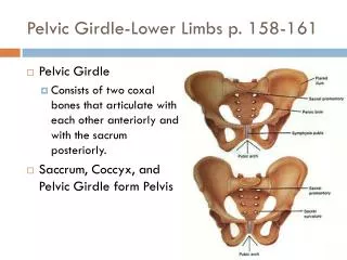
Hip Bone • The ilium, ischium, and pubis form the hip bone • They meet one another at the acetabulum. The hip bones articulate with the sacrum at the sacroiliac joints and form the anterolateral walls of the pelvis; they also articulate with one another anteriorly at the symphysis pubis.
The ilium • is the upper flattened part of the bone, possesses the iliac crest. This can be felt • through the skin along its entire length; it ends in front at the anterior superior iliac spine and behind at the posterior superior iliac spine. • The iliac tubercle lies about 2 in. (5 cm) behind the anterior superior spine. Below the anterior superior iliac spine is a prominence, the anterior inferior iliac spine; a similar prominence, the posterior inferior iliac spine, is located below the posterior superior iliac spine. Above and behind the acetabulum, the ilium possesses a large notch, the greater sciatic notch
The ischium • The ischium is L shaped, possessing an upper thicker part, the body, and a lower thinner part, the ramus. • The ischial spine projects from the posterior border of the ischium and intervenes between the greater and lesser sciatic notches. The ischial tuberosity forms the posterior aspect of the lower part of the body of the bone. • The greater and lesser sciatic notches are converted into greater and lesser sciatic foramina by the presence of the sacrospinous and sacrotuberous ligaments
The pubis • The pubis can be divided into a body, a superior ramus, and an inferior ramus . • The bodies of the two pubic bones articulate with each other in the midline anteriorly at the symphysis pubis; the superior ramus joins the ilium and ischium at the acetabulum, and the inferior ramus joins the ischial ramus below the obturator foramen. The obturator foramen in life is filled in by the obturator membrane. • The pubic crest forms the upper border of the body of the pubis, and it ends laterally as the pubic tubercle. • On the outer surface of the hip bone is a deep depression, called the acetabulum, which articulates with the • almost spherical head of the femur to form the hip joint. • The inferior margin of the acetabulum is deficient and is marked by the acetabular notch. • The articular surface of the acetabulum is limited to a horseshoe shaped area and is covered with hyaline • cartilage. The floor of the acetabulum is nonarticular and is called the acetabular fossa.
Femur • The femur articulates above with the acetabulum to form the hip joint and below with the tibia and the patella to form the knee joint. • The upper end of the femur has a head, a neck, and greater and lesser trochanters. • Thehead forms about two thirds of a sphere and articulates with the acetabulum of the hip bone to form the hip joint. • In the center of the head is a small depression, called the fovea capitis, for the attachment of the ligament • of the head. Part of the blood supply to the head of the femur from the obturator artery is conveyed along this ligament and enters the bone at the fovea.
Femur • The neck, which connects the head to the shaft, passes downward, backward, and laterally and makes an angle of about 125° (slightly less in the female) with the long axis of the shaft. The size of this angle can be altered by disease. • The greater and lesser trochanters are large eminences situated at the junction of the neck and the shaft. Connecting the two trochanters are the intertrochanteric line anteriorly, where the iliofemoralligament is attached, and a prominent intertrochanteric crest posteriorly, on which is the quadrate tubercle
Femur • The shaft of the femur is smooth and rounded on its anterior surface but posteriorly has a ridge, the lineaaspera, to which are attached muscles and intermuscular septa. The margins of the lineaasperadiverge above and below. The medial margin continues below as the medial supracondylar ridge to the adductor tubercle on the medial condyle. • The lateral margin becomes continuous below with the lateral supracondylar ridge. On the posterior surface of the shaft below the greater trochanter is the gluteal tuberosity for the attachment of the gluteus maximus muscle. The shaft becomes broader toward its distal end and forms a flat, triangular area on its posterior surface called the popliteal surface
Femur • The lower end of the femur has lateral and medial condyles, separated posteriorly by the intercondylar notch. The anterior surfaces of the condyles are joined by an articular surface for the patella. The two condyles take part in the formation of the knee joint. Above the condyles are the medial and lateral epicondyles. The adductor tubercle is continuous with the medial epicondyle.
Foramina of the Gluteal Region • The two important foramina in the gluteal region are the greater sciatic foramen and the lesser sciatic foramen. • Greater Sciatic Foramen • The greater sciatic foramen is formed by the greater sciatic notch of the hip bone and the sacrotuberousand sacrospinous ligaments. It provides an exit from the pelvis into the gluteal region.
Greater Sciatic Foramen The following structures exit the foramen: • Piriformis • Sciatic nerve • Posterior cutaneous nerve of the thigh • Superior and inferior gluteal nerves • Nerves to the obturator internus and quadratus femoris • Pudendal nerve • Superior and inferior gluteal arteries and veins • Internal pudendal artery and vein
Lesser Sciatic Foramen • The lesser sciatic foramen is formed by the lesser sciatic notch of the hip bone and the sacrotuberousand sacrospinous ligaments. It provides an entrance into the perineum from the gluteal region. Its presence enables nerves and blood vessels that have left the pelvis through the greater sciatic foramen above the pelvic floor to enter the perineum below the pelvic floor. • The following structures pass through the foramen: • Tendon of obturator internus muscle • Nerve to obturator internus • Pudendal nerve • Internal pudendal artery and vein
ARTERIES OF THE GLUTEAL REGION • There are three arteries coming into the gluteal region through the greater sciatic foramen: • superior gluteal artery • inferior gluteal artery • internal pudendal artery • These arteries are branches of the internal iliac artery which lies inside the pelvis.
PATELLA • The patella, or knee cap, is a triangular sesamoid bone embedded in the tendon of insertion of the quadriceps femoris muscle. • The superior border of the patella is the base of the triangle, and lateral and medial borders descend to converge at the apex. • A part of the quadriceps tendon covers the anterior surface of the bone and is continued, as the patellar ligament, to the tuberosity of the tibia.
PATELLA • The patella articulates on its posterior side with the patellar surface of the condyles of the femur. The articular surface of the patella comprises a larger, lateral facet and a smaller, medial one. Lateral dislocation of the patella is resisted by the shape of the lateral condyle of the femur and by the medial pull of the vastusmedialis. Excision of the patella results in minimal functional deficiency. The patella ossifies from several centers, which appear during childhood.
THIGH MUSCLES Fascial compartments of the thigh : • Anterior (Extensor) compartment = front of thigh. • Medial (Adductor) compartment = medial thigh. • Posterior (Flexor) compartment = back of thigh.
THIGH MUSCLES 1. Anterior compartment: Contains muscles that mainly extend the leg at the knee joint • Sartorius, iliacus, psoas, pectineus, and quadriceps Femoris (vastuslateralis +vastusmedialis + vastusintermedius + rectus femoris) - All supplied by femoral nerve.
THIGH MUSCLES 2. Medial compartment: - Consists of muscles that mainly adduct the thigh at the hip joint. - Adductor (magnus + longus + brevis), Pectineus, Gracilis, obturator externus - All supply by Obturator nerve.
THIGH MUSCLES 3. Posterior compartment: • Contains muscles that mainly extend the thigh at the hip joint and flex the leg at the knee joint. • Hamstrings (Biceps femoris + Semitendinosus + Semimembranosus) • All supplied by sciatic nerve.
FEMORAL TRIANGLE • The femoral triangle is a triangular depressed area formed by the inguinal ligament superiorly, the Sartorius muscle laterally, and the adductor longus muscle medially. The triangle contains the femoral nerve and its branches, the femoral sheath, the femoral artery and its branches, the femoral vein and its tributaries, and the deep inguinal lymph nodes. • The triangle is an important arterial pressure point in cases of severe hemorrhage of the lower limb. Hernias frequently occur in this area.
FEMORAL TRIANGLE BOUNDARIES & CONTENTS: 1. Femoral artery, artery of lower limb, starts from external iliac artery and ends into popliteal artery. 2. Femoral vein. 3. Inguinal lymph nodes (receives lymph from lower limb & external genetalia). 4. Great saphenous vein, ends into femoral vein. 5. Femoral nerve.
THE HIP JOINT • Type: synovial, ball & socket joint. • Boney articulation = Acetabulum of hip bone + Head of femur. (Deepened by the labrum). • Fibrous capsule: encloses the head and neck of femur. • Stability: The stability is high with lower range of movements (compared to shoulder joint). 1. Boney factor: close fitting bones. 2. Fibrous capsule and ligaments are strong. 3. Powerful muscles around the joint.
THE HIP JOINT • Ligaments: • The iliofemoral ligament • The pubofemoral ligament • The ischiofemoral ligament • The transverse acetabular ligament. • The ligament of the head of the femur
THE HIP JOINT • MOVEMENTS OF HIP JOINT: The hip joint allows flexion, extension, abduction, adduction, circumduction, medial rotation, and lateral rotation of the thigh.
Fracture Neck of The Femur • Fracture neck femur causes cutting of blood vessels to the head of femur and it’s “death” leading to damage of hip joint.
Bones of the Leg • Tibia • Fibula
Tibia • The Tibia is the large weight-bearing medial bone of the leg. • It articulates with the condyles of the femur and the head of the fibula above and with the talus and the distal end of the fibula below. • It has an expanded upper end, a smaller lower end, and a shaft.
Tibia • At the upper end are the lateral and medial condyles, which articulate with the lateral and medial condyles of the femur and the lateral and medial menisci intervening. Separating the upper articular surfaces of the tibial condyles are anterior and posterior intercondylar areas; lying between these areas is the intercondylar eminence • The lateral condyle possesses on its lateral aspect a small circular articular facet for the head of the fibula.
Tibia • The shaft of the tibia is triangular in cross section, presenting three borders and three surfaces. • Its anterior and medial borders, with the medial surface between them, are subcutaneous. • The anterior border is prominent and forms the shin. • At the junction of the anterior border with the upper end of the tibia is the tuberosity, which receives the attachment of the ligamentum patellae. The anterior borderbecomesrounded below, where it becomes continuous with the medial malleolus. • The lateral or interosseous border gives attachment to the interosseous membrane. • The posterior surface of the shaft shows an oblique line, the solealline, for the attachment of the soleus muscle.
Tibia • The lower end of the tibia is slightly expanded and on its inferior aspect shows a saddle-shaped articular surface for the talus. • The lower end is prolonged downward medially to form the medial malleolus. The lateral surface of the medial malleolus articulates with the talus. • The lower end of the tibia shows a wide, rough depression on its lateral surface for articulation with the fibula.
Fibula • The fibula is the slender lateral bone of the leg . • It takes no part in the articulation at the knee joint, but below it forms the lateral malleolus of the ankle joint. • It takes no part in the transmission of body weight, but it provides attachment for muscles. • The fibula has an expanded upper end, a shaft, and a lower end.