Download
1 / 26
270 likes | 294 Vues
Explore the characteristics and functions of chromosomes in mitosis and meiosis, key phases, and how errors in DNA replication can lead to cancer. Learn about karyotyping and the process of creating egg and sperm cells. Understand the importance of genetic screening and how chromosomes determine cell division.

E N D
The Chromosomes and karyotypePhases of Mitosis and Meiosis Lecture 4
glossary • Chromatin - diffuse, long fibers of DNA with proteins attached • Single chromosomes - a chromosome which has not been duplicated (like ½ of an x) • Duplicated chromosomes - a chromosome which has been duplicated (a complete X) • Chromatid - one half of a duplicated chromosome, looks like a single chromosome • Centromere - a structure made of protein which holds the two halves of a duplicated chromosome together.
The Chromosomes Male= XY Female= XX
Characteristics of Chromosomes 1. Location and Function • Located in the center of cells called the nucleus • Each chromosomes features protein and a single DNA molecule. • The DNA wrapped around histones (spool-like proteins) • A key part of the process of accurately copying DNA and distributing in many cell divisions
Characteristics of Chromosomes2. Pairs (23) • Female • 46 chromosomes • Two X chromosomes • One of each pair of the autosomes and one X is of maternal origin • Male • 46 chromosomes • Different pattern of sex chromosomes (XY) • One of each pair autosomes and an X is maternal origin • The father contributes the Y and the remaining autosomes
Characteristics of Chromosomes 3. Child Gender • The mother always contributes an X chromosomes to her child • The father of the contributes either an X chromosomes or a Y chromosomes • As a result, the father is the parent who determines the sex of a child. Still, a child inherits some traits from his mother and other traits from his father.
Characteristics of Chromosomes 4. Autosomal Types • Reproductive cells such as eggs and sperm must have the right number of chromosomes in order to have offspring that develops correctly. • Example: Down Syndrome • 3 copies of chromosome 21 instead of two copies • Autosomal abnormality
karyotypes definition Karyotyping Uses Refers to a picture of individual’s chromosomes. A technique that uses a picture of an individual’s chromosomes to analyze and detect chromosomal abnormalities Use to detect fetal abnormalities Form of genetic screening conducted on potential parents.
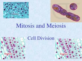
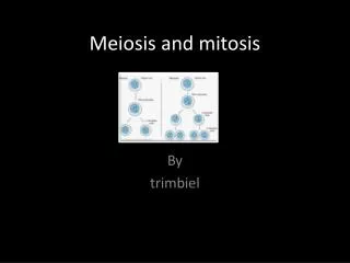
What is Mitosis? • Is the process, in the cell cycle, by which a cell duplicates into two genetically alike daughter cells. • Two identical cells from one original cell. • In mitosis, chromosomes in the cell nucleus are separated into two identical sets of chromosomes, each in its own nucleus. • Vital for tissue formation and maintenance
Phases/steps of mitosis • Prophase • Metaphase • Anaphase • Telophase
1. Prophase • The first stage of mitosis • Chromatin condenses to form duplicated chromosomes • The nuclear envelope breaks down (degenerate) • The spindle apparatus forms • The spindle attaches to the centromeres of the chromosomes and moves the chromosomes around during cell division
2. Metaphase • The second stage • The mitotic spindle moves the duplicated chromosomes so that they are lined up at the cell equator (middle of the cell) equidistant from the two poles (opposite ends) of the cell.
3. Anaphase • The third stage • Centromeres of duplicated chromosomes divide and split • The mitotic spindle pulls the sister chromatids separate from each other as they move to the opposite poles of the cell
4. Telophase • The fourth stage • Duplicated chromosomes decondense back into chromatin • The nuclear envelope begins to form around the chromosomes so that the two separate daughter nuclei are created • The mitotic spindle breaks down • Two daughter cells are produced • Diploid cells (2N)- 46 chromosomes same as the mother cells
Mitosis and Cancer • DNA, sometimes called a genetic blueprint, contains the hereditary material in nearly all organisms. • Two types of errors, or mutations, can occur when DNA is improperly copied: silent mutations, which have no impact on the DNA sequence, and "missense" mutations, which change a DNA sequence and often impact the associated function. • Missense mutations can multiply over time, leading to cell cycle disruption and formation of tumors -- cells that don't stop dividing. • Cancer occurs when the normal "checkpoints" regulating mitosis are ignored or overriden by a cancer cell, resulting in uncontrolled cell division.
What is Meiosis? • Types of cell division that creates egg and sperm cells • Ensures that humans have the same number of chromosomes in each generation. • A two step process that reduces the chromosomes number by half
Meiosis 1: • prophase 1, metaphase 1, anaphase 1, and telophase 1 • Meiosis 2: • prophase 2, metaphase 2, anaphase 2, and telophase 2
In the first meiotic division, the number of cells is doubled but the number of chromosomes is not. This results in 1/2 as many chromosomes per cell. • The second meiotic division is like mitosis; the number of chromosomes does not get reduced.
Disorders due to Meiosis Errors due during segregation (chromosomes segregates too many or few chromosomes in egg or sperm) • trisomy = an extra chromosome of a particular pair in each cell • monosomy = one chromosome fewer in each cell • For example • Trisomy 21 Down syndrome • cells have an extra copy of chromosome 21 (trisomy 21). • Turner syndrome • cells have only one X chromosome (monosomy X) and no Y chromosome