Download
1 / 17
270 likes | 766 Vues
Three-Dimensional Super-Resolution Imaging by Stochastic Optical Reconstruction Microscopy. Bo Huang, Wenqin Wang, Mark Bates , Xiaowei Zhuang. Science 319 , 810 (2008 ). Miyasaka Lab. Miyamoto Yoko. Contents. Introduction compare optical microscope and electron microscope

E N D
Three-Dimensional Super-Resolution Imaging by Stochastic Optical Reconstruction Microscopy Bo Huang, WenqinWang, Mark Bates, XiaoweiZhuang Science 319, 810 (2008) Miyasaka Lab. Miyamoto Yoko
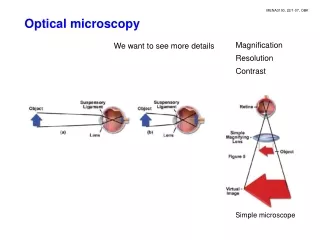
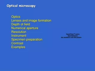
Contents • Introduction compare optical microscope and electron microscope localization method STORMstochastic optical reconstruction microscopy II. Experiments ,results and discussion Three- dimensional STORM Evaluation of spatial resolutionl of 3D STORM 3D STORM imaging of microtubules in a cell 3D STORM imaging of clathrin-coated pits in a cell III. Summary
STORM stochastic optical reconstruction microscopy Not need drying liquid living cell Super resolution microscopy Several tens of nm
STORM stochastic optical reconstruction microscopy Localization method Single molecule tracking X0 = 274.03 +/- 0.0339 pixel Y0 = 148.17 +/- 0.0351 pixel Actual precision of tracking ~several nm
STORM stochastic optical reconstruction microscopy High-resolution imaging technique Each molecule spatial resolution 1 nm diffractionlimit The overall image is then reconstructed from the fluorophore positions obtained from multiple imaging cycles.
STORM stochastic optical reconstruction microscopy For STORM , photo-switchable fluorophore is needed. Reporter a photo-switchable “reporter” fluorophorethat can be cycled between fluorescent and darkstates. Activator “activator” facilitates photo-activation of the reporter. Combinatorial pairing ofreporters and activators allows the creation of probes with many distinct colors. Alexa647633nm→ off state 532nm→ on state Cy3
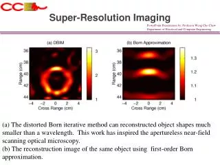
3D STORM 2D Recently, the diffractionlimit has been surpassed and lateral imaging resolutions of 20 to 50 nm have been achieved by several “super-resolution” far-field microscopy techniques. conventional image STORM image 3D But, most organelles and cellular structures cannot be resolved without high-resolution imaging in all three dimensions. →use the astigmatism imaging method to achieve 3D STORM imaging
3D STORM Y Focusing lens X: focus; Y: focus Isotropic intensity distribution X Z X: focus; Y: defocus Anisotropic intensity distribution Y Cylindrical lens Apparently Isotropic intensity distribution X Z X: defocus; Y: focus Anisotropic intensity distribution
3D STORM above focused in the y direction than in the x ellipsoidal with long axis along x the image appeared round the average focal plane below focused in the x direction than in the y ellipsoidal with long axis along y an elliptical Gaussian function h :the peak height b :the background (x0, y0) :the center position of the peak wx,wy :stand for the widths of the image in the x and y directions
3D STORM The wxand wyvalues as a function of z w0: the PSF width when a molecule is at the focal plane c : the offset of the x or y focal plane from the average focal plane d : the focus depth of the microscope A and B : coefficients of higher order terms to correct for the non-ideality of the imaging optics.
Three-dimensional localization distribution of single molecules Histograms of the distribution in x, y, and z Localizations from 145 clusters 22 nm 9 nm 11 nm Full width at half maximum value in x, y, and z 21 nm 26 nm 52 nm
3D STORM imaging of microtubules in a cell The 3D STORM image of the same area Conventional indirect immunofluorescence image
3D STORM imaging of microtubules in a cell fit to two Gaussians identical widths (FWHM = 66 nm) a separation of 102 nm (red curve)
3D STORM imaging of clathrin-coated pits in a cell V. I. Slepnev, P. De Camilli, Nat. Rev. Neurosci. 1, 161 (2000). Sequential stages in clathrin-mediated endocytosis at the presynaptic terminal
3D STORM imaging of clathrin-coated pits in a cell microtubule network in green monkey kidney epithelial (BS-C-1) cells Magnified view of two nearby CCPs in 2D STORM Conventional direct immunofluorescence image The 2D STORM image of the same area An x-y cross section (50 nm thick in z) of the same area the ring-like structure
3D STORM imaging of clathrin-coated pits in a cell (F) x-y cross sections (each 50 nm thick in z) (G) x-z cross sections (each 50 nm thick in y) of a CCP an x-y and x-z cross section presented in 3D perspective showing the half-spherical cage-like structure of the pit.
Summary 3D STORM determine both axial and lateral positions of individual fluorophoreswith nanometer accuracy . The image of each fluorophore simultaneously encodes its x, y, and zcoordinates, no additional time was required to localize each molecule in 3D STORM as compared with 2D STORM imaging. 3D STORM experiments demonstrate the ability to resolve nanoscopic features of cellular structures with molecular specificity under ambient conditions.