Optical Microscopy
Optical Microscopy. Widefield Microscopy - Brightfield, Darkfield, DIC, Phase Contrast, Fluorescence … Total Internal Reflection (TIR and TIRF) Microscopy Confocal Microscopy - fluorescence and reflection. Multiphoton or Nonlinear Microscopy Nearfield Microscopy (NSOM) 4-Pi Microscopy


Optical Microscopy
E N D
Presentation Transcript
Optical Microscopy • Widefield Microscopy - Brightfield, Darkfield, DIC, Phase Contrast, Fluorescence … • Total Internal Reflection (TIR and TIRF) Microscopy • Confocal Microscopy - fluorescence and reflection. • Multiphoton or Nonlinear Microscopy • Nearfield Microscopy (NSOM) • 4-Pi Microscopy • STED Microscopy (STimulated Emission Depletion) • Structured illumination microscopy (SIM) and saturated structured illumination microscopy (SSIM) • Selective plane illumination microscopy
Optical Sectioning in Biological Microscopy Fluorescently labeled sea urchin eggs Conventional light microscopy doesn’t work well on thick (> few microns) specimens Fixation and Physical Sectioning Widefield Fluorescence Deconvolution Methods (Computational) Widefield Fluorescence Live specimens Confocal Microscopy Confocal Aperture Multiphoton Microscopy nonlinear processes
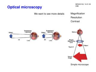
Laser scanning microscopy The focused laser is raster scanned across the sample and the fluorescence is detected, amplified and digitized. Objective lens
Confocal Microscopy produces optical sections by excluding light from outside of the focal plane. Fluorescence emission excitation
Two-Photon, Multiphoton or Nonlinear Microscopy uses nonlinear optical processes to create contrast and obtain optical sectioning. The two most common nonlinear processes are Two photon fluorescence and second harmonic generation (SHG):
In vivo imaging - example: transgenic mouse models of Alzheimer's disease. 3D projection of b amyloid plaque stained with Thio-S, excitation at 760 nm. Christie, R. H., Bacskai, B. J., Zipfel, W. R., Williams, R. M., Kajdasz, S. T., Webb, W. W. & Hyman, B. T. (2001) J Neurosci 21, 858-64. Bacskai, B. J., Kajdasz, S. T., Christie, R. H., Carter, C., Games, D., Seubert, P., Schenk, D. & Hyman, B. T. (2001) Nat Med 7, 369-72.
Transgenic mouse models of ovarian cancer based on p53 and Rb inactivation Histology MPM Intrabursal injections of AdCre into mice carrying conditional p53, Rb1 or both alleles results in ~100% epithelial neoplasms With Alexander Niktitin’s laboratory, Biomedical Sciences
Is it possible to use nonlinear laser scanning microscopy to image a ~cm field of view* as an aid, for example, to better define tumor borders? Advantages may be: 1. Better 3D view. 2. Maximum optical resolution could still be on the order of ~4 microns and the system would be able to zoom to the cellular level. 3. Ability to excite both targeted contrast agents (example – 5-ALA -> protoporphyrin IX) and use intrinsic signals for an overall tissue view. Disadvantages: 1. A more complex instrument. 2. Since it would be used with a conventional surgical microscope, image registration may be difficult to achieve. *Typical field of view in a laser scanning (confocal or multiphoton) microscope is ~0.5 x 0.5 mm
Intraoperative Fluorescence microscope from Zeiss – OMPI Pentero Glioblastoma IV under white light and under BLUE 400 illuminationWalter Stummer, M.D., University of Düsseldorf, Düsseldorf, Germany
Multiphoton imaging with a 2x lens (0.14 NA) - field of view is 7 mm (movie is of the word “Cornell” in 12 pt font from my business card)
Ascites tumor model (transformed p53/Rb ovarian epithelial cells injected IP) Widefield fluorescence image (cells also express GFP) tumor White light image of small (~3 mm diameter) metastasis on small instestine
Two color multiphoton imaging of tumor on the small intestine in an ascites tumor model 7 mm