Download

1 / 34
350 likes | 588 Vues
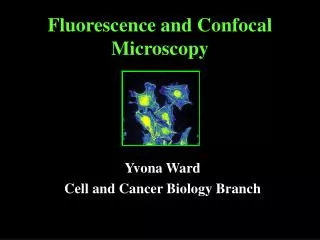
THREE DIMENSIONAL RECONSTRUCTION OF EXOSKELETON BY CONFOCAL MICROSCOPY. TROCHANTER OF FRONT LEG. Confocal microscopy: DAVID NEFF LAURA QUIMBY FAITH FRAZIER. CONVERTING CONFOCAL MODELS OF EXOSKELETON TO MODELS FOR FINITE ELEMENT ANALYSIS. TROCHANTER OF HINDLEG.

E N D
THREE DIMENSIONAL RECONSTRUCTION OF EXOSKELETON BY CONFOCAL MICROSCOPY TROCHANTER OF FRONT LEG Confocal microscopy: DAVID NEFF LAURA QUIMBY FAITH FRAZIER
CONVERTING CONFOCAL MODELS OF EXOSKELETON TO MODELS FOR FINITE ELEMENT ANALYSIS TROCHANTER OF HINDLEG CLAY FLANNIGAN, CASE WESTERN
PEGLEG EXPERIMENTS: PHYSIOLOGY AND FINITE ELEMENT ANALYSIS STUDIES
PEG LEG TESTS ON HINDLEG Clay Flannigan, Case Western FORCE EXTEND FORCE FLEX P RESULTS - RECEPTORS SIGNAL FORCES AS AN ARRAY - SOME GROUPS ARE EXCITED BY EXTERNAL LOAD AND MUSCLE CONTRACTIONS AND CAN PROVIDE POSITIVE FEEDBACK
TROCHANTERAL GROUP 3 SENSILLA ENCODE FORCED FLEXIONS: PHYSIOLOGY ABLATION EXPERIMENTS
STRAINS PRODUCED BY EXTENSOR MUSCLE CONTRACTIONS: SOME GROUPS CAN MEDIATE POSITIVE FEEDBACK
STRAINS DURING STANCE PHASE:S. Ramasubramanian, G. Nelson, Case Western Fy Fz Fx Femur and Tibia Attached to Trochanter - FT Joint Angle from Kinematic Studies, Forces applied at Levels (Fx, Fy, Fz) for early, middle and late stance.(Kinematic Data from J. Watson, R. Ritzmann; Force Data from R. Full)
STRAINS DURING MIDSTANCE PHASE:S. Ramasubramanian, Case Western LEG LOADING RESULTS -ARRAY ENCODES FORCE DIRECTION (FORCE POSTERIOR, FLEXION) -ARRAY ENCODES FORCE MAGNITUDE
TROCHANTERAL FORCE SENSORS AS LOAD CELL: Fx, Fy and Fz INDEPENDENTLY VARIED ARRAY CAN DETECT INCREASES IN FORCE VECTORS
Summary: ENCODING PROPERTIES OF CAMPANIFORM SENSILLA- signal level of load rate of change of force, provide active signal for sudden decreases MODELING THROUGH FINITE ELEMENT ANALYSIS - trochanteral campaniform sensilla encode forces as a array; some groups provide positive feedback in limited ranges of joint angles LEG DESIGN/ANATOMY DETERMINES SPECIFICITY OF AFFERENT INPUTS
FEA Modeling Studies:Strain in Exoskeleton 1- Exoskeleton is a tubular structure that is rigid (modulus =3160 N/mm^2); fixed beam analysis is useful 2- Receptors located on proximal ends of segments and are subject to large bending forces when end of leg is pushed against substrate • 3- Strains also result from muscle contractions: • Muscles with insertions near receptors produce strains; locally distribution depends upon shape • Muscles located close to body can produce bending along length of leg TIBIAL EXTENSOR MUSCLE TIBIAL SENSILLA TROCHANTERAL SENSILLA TROCHANTERAL EXTENSOR MUSCLE
Three Dimensional Reconstruction of Exoskeleton by Confocal Microscopy Converted to FEA Model CLAY FLANNIGAN, ROGER QUINN CASE WESTERN RESERVE FEA MODEL Confocal microscopy: DAVID NEFF LAURA QUIMBY FAITH FRAZIER
FORCE RECEPTORS ONTROCHANTER The trochanteral segment contains the largest number of strain sensing campaniform sensilla in the leg. These receptors have been characterized physiologically and their responses have been modeled by Finite Element Analysis. The trochanteral receptors can provide precise information about the magnitude, direction and rate of forces applied to the femur.
Insect-Like Hexapods • Robot II - “stick insect” • 6 symmetric 3 DOF Legs - 18DOF • Locomotion controller uses network of inter-leg influences • Able to walk over rough and unstable terrain Robot II • Robot III - cockroach • Pneumatic actuation • 24 DOF: 5 front, 4 middle, 3 rear • Currently shows high power (30lb payload) and robust posture control Robot III
Cockroach Anatomy • 5 leg segments: coxa, trochanter, femur, tibia, tarsus • CT, FT are 1 DOF, hinge-like and articulate about 2 condyles • Large muscles of the coxa insert on the trochanter • Trochanter is heavily loaded during stance Coxa Tibia Tarsus Femur Trochanter Cockroach Leg Muscle Definition
Trochanter • Approximately 2.5 mm in length for the adult P. americana • Outer surface is smooth; inner surface has variable topology • Coxa attaches via 2 condyles on the anterior and posterior sides • Femur attaches via 2 condyles on the distal anterior surface Dorsal Coxal Condyles Femur Distal Proximal Femoral Condyles y Trochanter x Ventral Anterior view trochanter
Campaniform Sensilla • Campaniform Sensilla (CS) are cuticular caps in the exoskeleton innervated by a single nerve • Ellipsoidal with long-axis length from 6 to 24 mm • Have been shown to respond to cuticular compressive strains perpendicular to the long-axis (short axis strains) • Directionally sensitive Cuticle Cap Dendrite Sensory Neuron Axon Campaniform sensilla structure
TrochanteralCampaniform Sensilla • CS found in 6 groups on the leg (10-15 in a group) • Within groups the CS are similarly oriented • 4 groups on the trochanter - 3 on the anterior 1 on the posterior • Notice the ridge structures and ring of the interior surface Group 3 Group 4 Group 2 Anterior campaniform sensilla groups Group 1 Posterior campaniform sensilla group
Confocal Modeling(Zill Lab) • Confocal microscope used to optically section the trochanter • Irradiation of the specimen with UV Light causes the cuticle to fluoresce • Microscope indexes its focal plane through the trochanter • Result: A series of bitmap images, when combined using specialized software, creates 3-D model of the trochanter Confocal Sections
Meshed Trochanter Thickness Scale: red 6mm blue 265mm Anterior Posterior
Forced Flexion • Von Mises strain: Anterior Posterior
STRUCTURE AND LOCATION OF CAMPANIFORM SENSILLA FUNCTION LIKE STRAIN GAUGES DETECT STRAIN IN EXOSKELETON - COMPRESSIONS PERPENDICULAR TO CAP LONG AXIS EXCITE RECEPTOR
TROCHANTERAL CAMPANIFORM SENSILLA:IMAGING BY CONFOCAL MICROSCOPY 1) OCCUR AS FOUR GROUPS (Gp1-4) 2) EACH GROUP HAS CONSISTENT ORIENTATION 3) GROUPS ARE ASSOCIATED WITH INTERNAL BUTTRESSES OR THICKENINGS OF EXOSKELETON OUTER SURFACE INNER SURFACE diI OUTFILL CONFOCAL IMAGES OF CUTICLE
THREE DIMENSIONAL RECONSTRUCTION OF EXOSKELETON BY CONFOCAL MICROSCOPY Confocal microscopy: DAVID NEFF LAURA QUIMBY FAITH FRAZIER TROCHANTER OF FRONT LEG
ACCURACY CONFIRMED BY SECTIONING MODEL/ORIGINAL TROCHANTER 3D RECONSTRUCTION SEM
CONFOCAL and FEA models Confocal model FEA model Overlay comparison
ACTIVITIES OF CAMPANIFORM SENSILLA ARE EVALUATED BY STRAINS AT LOCATION OF CUTICULAR CAPS - nodes corresponding to CS are located on the finite element model - Compressions (negative strain values) excite receptors Group 1, posterior trochanter Groups 2,3,4, anterior trochanter
FEA PEGLEG EXPERIMENT BEHAVIORAL PARADIGM PHYSIOLOGY PARADIGM ATTACH PEG LEG BEND FEMUR SENSORY RECORDING FEA PEGLEG APPLY FORCES TO ATTACHED FEMUR, POINTS OF MUSCLE INSERTIONS
PEG LEG TEST ON Blaberus Discoidalis FRONT LEG FORCE FLEX FORCE EXTEND Group 3 short axis strain Group 4 short axis strain RESULTS: Campaniform Sensilla - Function as a strain gauge array signals forces in all directions of load - Can provide positive feedback
Fz Fy Fx FINITE ELEMENT MODELLING OF STRAINS DURING WALKING High speed video: Adam Noah Angela Ridgel Mid-stance Late stance
SENSORY ACTIVATION PATTERNS DUE TO GROUND REACTION FORCES DURING STANCE • Groups 1 and 4 show excitation due to ground reaction forces throughout stance. • These sensilla groups could be responsible for signaling that leg is on substrate and bearing load.