Fluorescence and Confocal Microscopy
Fluorescence and Confocal Microscopy. Yvona Ward Cell and Cancer Biology Branch. OUTLINE. Immunofluorescent Staining Conventional Fluorescent Microscopy Confocal Microscopy Applications. Immunofluorescent Staining. Immunofluorescent staining makes use of antibodies to locate and

Fluorescence and Confocal Microscopy
E N D
Presentation Transcript
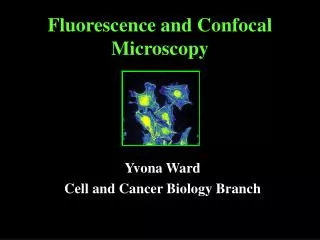
Fluorescence and Confocal Microscopy Yvona Ward Cell and Cancer Biology Branch
OUTLINE • Immunofluorescent Staining • Conventional Fluorescent Microscopy • Confocal Microscopy • Applications
Immunofluorescent Staining Immunofluorescent staining makes use of antibodies to locate and identify patterns of protein expression in cells. Primary antibody binds to antigen. Antibody-antigen complex is bound by a secondary antibody conjugated to a fluorochrome. Upon absorption of high energy light, the fluorochrome emits light at its own characteristic wavelength (fluorescence) and thus allows detection of antigen-antibody complexes. Suitable for: 1. frozen, non-fixed tissues and ethanol fixed tissues 2. paraformaldehyde-fixed or methanol/acetone-fixed cells
Basic Staining Technique • Immunofluorescent Cell Staining • Incubate cells with a blocking solution to minimize non-specific staining • Incubate cells with a polyconal or monoclonal antibody specific for the protein of interest. • Incubate cells with a secondary antibody directed against the primary antibody. • The secondary antibody must be conjugated to a fluorochrome PRIMARY ANTIBODY SECONDARY ANTIBODY FLUOROCHROME ANTIGEN • Cell Preparation • Culture cells on a glass coverslip in a 24-well plate. Cells may be transfected directly on the • coverslip. • Fix cells using paraformaldehyde or methanol/acetone and then wash them 3 times in PBS • Cell Permeabilization • Incubate fixed cells in 1% Triton X-100 in PBS+0.02%BSA for 2 minutes at room temperature. • Wash the cells 3 times with PBS.
Giannakakou et al., Nature Cell Biology,2000 PRIMARY ANTIBODY sheep anti-p53 polyconal SECONDARY ANTIBODY Texas Red conjugated anti-sheep PRIMARY ANTIBODY mouse anti-a tubulin monoclonal SECONDARY ANTIBODY FITC conjugated anti-mouse
Direct Staining of Cell Structures Organelle Probes Mitochondria MitoTracker mitochondrial membrane potential Lysosomes LysoTracker hydrolytic activity of enzymes ER and Golgi Lectin conjugates lipid composition Other Probes Stressfibers Phalloidin-conjugaes bind F-actin Nuclei DAPI binds to minor groove of ds-DNA
MitoTracker-Orange CMTMRos 4 4’,6-diamidino-2-phenylindole (DAPI) TRAP1 Mouse Monoclonal + Goat anti-Mouse-FITC Felts et al., JBC, 2000
centrosomes microtubules Anti-pericentrin PoAb Goat anti-mouse-FITC Anti-tubulin MoAb Goat anti-mouse-Rhodamine DAPI nucleus MERGE
Stress Fibers Focal Adhesions Anti-vinculin MoAb Goat anti-mouse-FITC Rhodamine-Phalloidin
Translocation of mutated protein to the mitochondria deep red-mitotracker GFP-fusion protein MERGE
Use of Biotinylated Antibodies PRIMARY ANTIBODY SECONDARY ANTIBODY ANTIGEN FLUOROCHROME BIOTIN STREPTAVIDIN Streptavidin is a bacterial protein that specifically binds biotin. This interaction may be used to label cellular components.
Rabbit anti-Factor H Biotinylated Goat anti-Rabbit Streptavidin-FITC Guinea Pig anti-Insulin PoAb Donkey anti-Guinea Pig-Cy5 Mouse anti-Glucagon MoAb Goat anti-Mouse-Rhodamine Martinez et al., J. Endocrinol., 2001
Conventional Fluorescent Microscopy
Confocal Microscopy Core Inverted Scope Upright Scope
Preparation of Stained Specimens For Microscopy Specimen Mounting • In order for the stained specimen to be visualized on a fluorescent • microscope, it needs to be mounted onto a slide using an appropriate • mounting medium. • Mounting medium is usually a PBS/Glycerol mix and is commercially • available. • Biomeda Corporation Aqueous Mounting Medium • Molecular Probes SlowFade
Specimen Photobleaching • One of the major problems in microscopic examination of fluorescent specimens is the tendency of fluorochromes to lose fluorescence upon excitation by a high energy light source. • Free radicals generated during fluorochrome excitation are responsible for this quenching or photobleaching. • Various chemical agents that scavenge free radicals may be added to the mounting medium to preserve specimen brightness. • Sigma trans-pyridine-2-azo-p-dimethylaniline (PADA)
Fluorescence Molecules absorbing the energy of electromagnetic radiation will jump to a higher energy level. When certain excited molecules return to the ground state they emit radiation. This phenomenon is known as fluorescence. Fluorescent molecules are known as fluorochromes or fluorophores.
Fluorochrome Absorption Emission Cascade Blue 400 420 Fluorescein 494 518 Rhodamine 570 590 Texas Red 595 615 Cy5 650 670 Absorption Spectra of Fluors Commonly Conjugated to Secondary Antibodies
Fluorescence Microscopy • Since the molecules used for immunofluorescence emit light in the • visible range, it is possible to detect them with a microscope. • A mercury lamp is used to illuminate the sample with UV light through the objective lens. A dichroic mirror reflects short l and transmits longer l. • The fluorescence emitted from the sample passes back through this mirror, but the UV light does not. • An excitation filter in front of the mirror will control the excitation wavelength. • An emission filter in front of the eyepiece will control the wavelength of the emitted light. eyepiece excitation filter emission filter * Hg dichroic mirror specimen
Numerical Aperature (NA) A solid cone of light that hits the specimen Lenses with a high NA have a short working distance but, allow more light to be captured from the specimen. Example: Phase contrast lens low NA long working distance High resolution 100x high NA short working distance
UV 351,364nM Argon 488nM HeNe I 543nM HeNe 2 633nM CCBB Confocal Core Facility (1999-2006) Zeiss LSM510 with 4 color capability Building 37 Room 1035
What is Confocal Microscopy? Laser Scanning Confocal Microscopy Confocal Scanning Laser Microscopy Confocal microscopy is a powerful tool for generating high-resolution images and 3-D reconstructions of a specimen. In confocal microscopy a laser light beam is focused onto a fluorescent specimen through the objective lens. The mixture of reflected and emitted light is captured by the same objective and is sent to the dichroic mirror. The reflected light is deviated by the mirror while the emitted fluorescent light passes through a confocal aperature (pinhole) to reduce the “out of focus” light. The focused light then passes through the emission filter and proceeds to the photomultiplier. In order to generate an entire image, the single point is scanned in an X-Y manner as the laser focus is moved over the specimen.
The LSM 510 • To the Specimen • optical fibers • main dichroic beam-splitter • scanner mirrors • scanning lens • From the Specimen • optical fibers • main dichroic beam-splitter • (7,8,9) secondary dichroics • (10) pinhole diaphragm • (11) emission filters • (12) photomultipliers
Why is Confocal Microscopy Better? 1. More Color Possibilities Because the images are detected by a computer rather than by eye, it is possible to detect more color differences.
Insulin-Cy5 CRLR-FITC Overlay Glucagon-Rhodamine
Why is Confocal Microscopy Better? 2. Less Cross Talk In most applications, fluorochromes have overlapping emission spectra. Hence, the emission signals cannot be separated completely into different detection channels resulting in “bleed through” or cross talk. However, if the fluorochromes have distinct excitation spectra, the fluorochromes can be excited sequentially using one excitation wavelength at a time. This is only possible with confocal systems that offer the multitracking feature.
Standard Microscopy Multitracking Brain Slice nerve fibers (FITC) cell nuclei (propidium iodide) Courtesy Dr. Schild, University of Gottingen
Why is Confocal Microscopy Better? 3. Optical Sectioning of Objects Without Physical Contact Zebra fish embryo wholemount Neurons (green) Cell adhesion molecule (red) Monika Marks, Martin Bastmeyer University of Konstanz
Formation of Acini in a 3-D Matrigel Matrix Three-dimensional culture of MCF10A mammary epithelial cells on a reconstituted basement membrane leads to the formation of polarized, growth arrested acini-like spheroids that recapitulate several aspects of glandular architecture in vivo. Introduction of oncogenes into MCF10A cells results in distinct morphological phenotypes
Empty vector b-catenin DAPI MERGE Ras V12
Why is Confocal Microscopy Better? 4. Three-Dimensional Reconstruction of Specimen 3D shadow projection Tight junctions (red) Cytoskeletal structures (green) Prof. Wunderli-Allenpach ETH, Zurich
Animated 3-Dimensional Reconstruction Laser Scanning Microscopy LSM510 3D for LSM www.Zeiss.com
Animated 3-Dimensional Reconstruction Mitosis www.Zeiss.com
Why is Confocal Microscopy Better? 5. Improved Resolution Rat Cerebellum Astrocytes (green) Mn dismutase (red) Jorg Lindeman University of Magdeburg
Applications • Colocalization • Live Cell Imaging • FRAP/FLIP • GFP-Fusion • 3. FRET
Colocalization of Proteins Colocalization of up to 4 different proteins Colocalization does not mean interaction Decreased cross talk with multitracking feature
Colocalization of insulin and calcitonin receptor-like receptor Insulin-Cy5 CRLR-FITC Glucagon-Rhodamine
Colocalization a-tubulin p53 Proteins may colocalize but not necessarily interact
Fluorescence Resonance Energy Transfer The high resolution of a confocal microscope allows us to study the physical interaction of protein partners.
What is FRET? FRET is the non-radioactive transfer of photon energy from an excited fluorophore (the donor) to another fluorophore (the acceptor) when both are located within close proximity (1-10nm). Using FRET one can resolve the realtive proximity of molecules beyond the optical limit of a light microscope to reveal (1) molecular interactions between two protein partners, (2) structural changes within one molecule (eg. enzymatic activity or DNA/RNA conformation), (3) ion concentrations using special FRET-tools like the CFP-YFP cameleon CFP is excited by light and emits light CFP is more than 10nm from YFP YFP is not excited and does not emit light No FRET Signal CFP is excited by light and emits light CFP is in close proximity to YFP YFP emits light FRET Signal
The Principle of FRET An excited fluorophore (donor) transfers its excited state energy to a light absorbing molecule (acceptor). This transfer of energy is non-radioactive due primarily to a dipole-dipole interaction between donor and acceptor. There are only certain pairs of fluorophores suitable for FRET experiments since, besides other prereqisites (eg. dipole orientation or sufficient fluorescence lifetime), the donor emission spectrum has to overlap the excitation spectrum of the acceptor. Known FRET pairs are CFP/YFP, BFP/GFP, GFP/Rhodamine, FITC/Cy3. Energy Diagram of CFP/YFP FRET: CFP donor is excited but most of its energy does not result in cyan emission. Instead, It is transferred to the YFP acceptor. Thus Resulting emission is mostly yellow.
FRET Region of interest Two channel (CFP,YFP) time series Two channel (CFP,YFP) time series
Confocal Microscopy is a powerful tool for studying signaling mechanisms Yvona Ward Building 37 Room 1066 301-594-2645 yward@helix.nih.gov