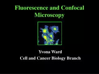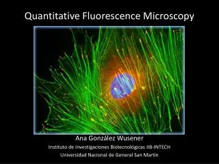
Fluorescence Microscopy
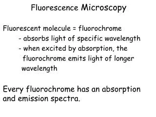
Fluorescence Microscopy. Test your prediction skills. Hoechst stain. The structure of these stains causes them to slide in between the bases of DNA and fluoresces blue. What structure of the cell would you predict would be stained?. Human cheek cell. We’ll come back to the green.


Fluorescence Microscopy
E N D
Presentation Transcript
Fluorescence Microscopy Test your prediction skills.
Hoechst stain • The structure of these stains causes them to slide in between the bases of DNA and fluoresces blue. • What structure of the cell would you predict would be stained?
Human cheek cell We’ll come back to the green.
Connective tissue cells – fibroblasts We’ll come back to the red and green.
Bull sperm The heads of sperm are filled completely with this structure. We’ll come back to the green.
DiOC6associates with lipids and produces a green color What subcellular structures are lipid based?
Oxidation revealer red Concentrated in the organelle that uses oxygen to generate ATP
Oxidation revealer green Same characteristics as oxidation revealer red, but a different color fluorescence
oxidation revealing greensame structure, highly elongated shape Fathead minnow cell
acid loving red Accumulates in organelles with an acid pH, useful for breaking down wastes
Canine kidney cells We’ll come back to the green, which is a different dye from the one you’ve seen already.
BODIPY ceramide(green) Ceramides are molecules taken up and metabolized by the organelle responsible for for transporting lipids and proteins to the cell membrane
Phalloidin green Binds to filamentous actin
Phalloidin red Binds to filamentous actin
Bovine pulmonary artery endothelial cells – the cells lining the interior of the major vessel bringing blood into the lungs. In this picture, the green is a microtubule stain. The red is phalloidin.