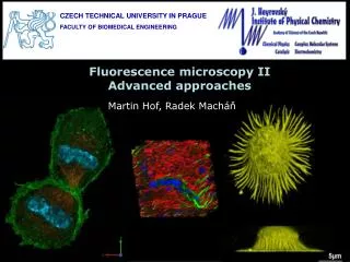
Fluorescence Microscopy
Fluorescence Microscopy. G.A.M.E.S. 2008 Bioengineering Camp. Light travels through many important parts of the microscope. Light of one wavelength comes from microscope light Travels through excitation filter Reflects off dichromic mirror (down) Hits sample Sample emits light (up)


Fluorescence Microscopy
E N D
Presentation Transcript
Fluorescence Microscopy G.A.M.E.S. 2008 Bioengineering Camp
Light travels through many important parts of the microscope. • Light of one wavelength comes from microscope light • Travels through excitation filter • Reflects off dichromic mirror (down) • Hits sample • Sample emits light (up) • Travels through dichromic mirror • Travels through emission filter • Is seen by eye • (page 6)
The fluorescence microscope diagram shows these light paths.
Actin is stained by rhodamine phalloidin. • Reminder: rhodamine phalloidin is two different molecules that work together. • Rhodamine – fluorescence • Phalloidin – attaches to actin • Green light makes rhodamine molecules move around; they release red light at a lower energy (longer wavelength) • What you can expect to see • Black background with red stress fibers visible • No green
Photobleaching makes the fluorescence fade. • If any fluorescent sample is left exposed too long to light, the fluorescence will fade. • Protect sample from light after adding rhodamine phalloidin – cover with aluminum box. • Results will not be as good after photobleaching.
GFP makes transfected cells glow green. • Reminder: GFP is made by cells that have taken up the new DNA. • Just like rhodamine, the GFP molecules move around when specific light is used. • Shine on blue, glow green.
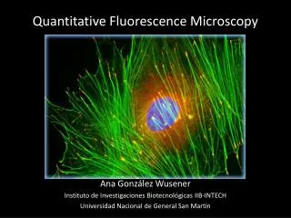
Multi-colored fluorescence images are hard to make. • Must change the color of light from the microscope • Must layer each picture – no single picture looks like this --> • Blue is the nucleus • Red is actin • Green is golgi • Pink is mitochondria
Emitted light is always a different color than the original one. GFP (Transfection) Actin Staining Purple Green Orange Blue Yellow Red