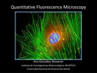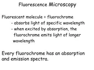
Fluorescence Microscopy
Fluorescence Microscopy. Chelsea Aitken Peter Aspinall. Advantages Over Light Microscopy. Resolution of light microscopy limited by Rayleigh Criterion If two objects cannot be seen as distinct structures, then they may be considered coincident in space


Fluorescence Microscopy
E N D
Presentation Transcript
Fluorescence Microscopy Chelsea Aitken Peter Aspinall
Advantages Over Light Microscopy • Resolution of light microscopy limited by Rayleigh Criterion • If two objects cannot be seen as distinct structures, then they may be considered coincident in space • Unable to determine whether there are molecular associations • Fluorescence microscopy can determine the distance between two molecules to 20 – 100 Å
Wide-Field Fluorescence Microscope • Molecule (fluorophore) absorbs a photon and then quickly reemits a lower energy photon • Change in energy allows us to filter out incident light • Uses epi-illumination • Light source goes into a filter cube and is reflected into the sample • Emission returns through same objective and filter cube • Because of its longer wavelength, it passes through the dichroic mirror and is read http://upload.wikimedia.org/wikipedia/commons/4/49/Dichroic_filters.jpg
Two-Photon Excited Microscopy • Simultaneous absorption of two photons causing the fluorophore to emit a higher energy (2x) photon • Simultaneous = ~ 10-18 s • However to generate the same number of two-photon events, the laser needs to be ~106times more powerful than for one-photon events • Use mode-locked (pulsed) lasers • Intensity at peak is great enough to cause two-photon events
Three-Photon Excited Microscopy • Three-photon events • Photon density needed is only ten times that needed for two-photon events • Useful to excite fluorophores that fluoresce at very short wavelengths (that can be difficult to produce) http://upload.wikimedia.org/wikipedia/commons/thumb/d/d3/MultiPhotonExcitation-Fig1-doi10.1186slash1475-925X-5-36.JPEG/1280px-MultiPhotonExcitation-Fig1-doi10.1186slash1475-925X-5-36.JPEG
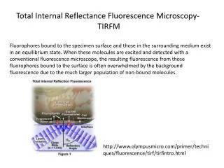
Total Internal Reflectance Fluorescence Microscopy (TIRFM) • Laser is pointed through one medium in contact with the medium of interest • Because of differences in refractive indices the light only reaches a short distance into the solution • Allows us to image cellular binding structures
4Pi-Confocal Microscopy • Uses two objective lens • One to illuminate • One to observe • This doubles the aperture angle making it possible to have an aperture angle of 4pi • Produces clearer more detail images • Can be further improved by combining this setup with standing wave microscopy
Stimulated Emission Depletion Microscopy (STED) • Fluorophoresin a small area are excited by a short pulse of light (200 fs) • Then fluorophores around this area are forced back to ground state by a second longer pulse (40 ps) • This creates a very sharp peak of fluorescence and increases resolution
Standing-Wave Illumination Fluorescence Microscopy (SWFM) • Two laser planes cross in the solution and creates an interference pattern • The nodes have spacing: • Theta (the angle between the two planes) can be varied to reduce the node spacing to • This allows it to have a better resolution than other methods
Fluorescence Resonance Energy Transfer (FRET) • A donor fluorophore is excited and then transfers its energy to an acceptor (if its close enough) through dipole-dipole interactions • Measures molecule interactions efficiency (ET) • R0 is the distance at which 50% of the energy is transferred • k2 – relative position of dipoles • J(λ) – integral of the overlap between the acceptor and donor spectra • QD– quantum yield of the donor • This method can be used as a quantum ruler (solve for R,) since ET can be measured and R0 can be calculated
Applications of FRET • Very useful for studying how DNA’s form changes when introduced to certain proteins • Label both ends of DNA and then can measure how the distance changes • Much better at measuring changes in distance than absolute distance • Very good spectroscopic ruler for 20 – 100 Å range • However cannot detect dynamic events
Green Fluorescent Protein (GFP) • Not all molecules fluoresce, so to use fluorescence microscopy they need to be fluorescently labeled • Dye molecules have to bind to specific location and not interfere with the reaction being monitored or the cell in general • Use GFP from Aequoria Victoria (type of jellyfish)
Pros/Cons of GFP • Pros • When expressed is spontaneously fluorescent • Doesn’t interfere with bound protein function • Can target specific organelles • Mutants have varying fluorescent properties • Cons • Limited sensitivity • Very large -> limits resolution • Can undergo color changes from irradiation independent from FRET • Takes hours to fold into its fluorescent shape
Applications of GFP • Conformational Sensor • Uses FRET between GFP and BFP to measure distance • Position varies as the bound protein undergoes structure changes • Can use reemitted wavelength of light to determine distance • Cellular Reporter • Can image living cells in vitro • Picture uses CFP (cyan) and YFP (yellow) • Can again use FRET principles with two different dyes • Measure conformational changes when two complexes interact
Fluorescence Lifetime Imaging Microscopy (FLI) • Image the fluorescence lifetime of all fluorophores in a sample • Can image live cells this way • Also possible to calculate FRET efficiencies at every pixel: • Where τD and τDA are the donor and acceptor lifetimes respectively • Can use this technique to visualize the locations of GFPs in a living cell in real time • Allows us to see cellular events in real time
Sources • Serdyuk, Igor N., Nathan R. Zaccai, and Joseph Zaccai.Methods in Molecular Biophysics: Structure, Dynamics, Function. New York: Cambridge University Press, 2007. Print.