Download
1 / 20
210 likes | 375 Vues
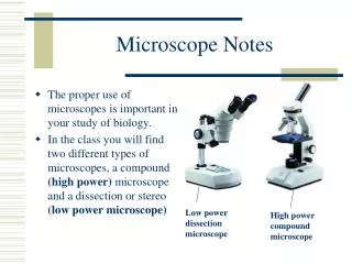
GFP Fluorescence/ Confocal Microscope. Kimberly Ching ABE Workshop Summer 2007. Fluorescent Microscope. Multiple Labeling ID specific organelles Necrotic vs. apoptotic cells Verify cell membrane permeability Locate antigen-specific molecules.

E N D
GFP Fluorescence/Confocal Microscope Kimberly Ching ABE Workshop Summer 2007
Fluorescent Microscope • Multiple Labeling • ID specific organelles • Necrotic vs. apoptotic cells • Verify cell membrane permeability • Locate antigen-specific molecules
Light from the excitation filter excites the fluorochoromes to a higher energy state From the high state it declines slowly releasing energy Transition between absorption & emission Alexander Jablonski Diagram
Excitation and Emission • Stokes Shift/Law • Florescence emission wave length is longer • Excitation wave length is shorter
Light from excitation filter thru objective lens; light absorbed Light emitted goes back thru objective lens, barrier filter, then detector Light Path
Fluorochromes • DAPI: blue dye, 4',6-diamidino-2-phenylindole, binds to DNA excited with UV, absorption max358nm, emission max 461nm • GFP: Green Fluorescence Protein, Aequorea victoria, absorption 488nm, emission 509nm • Fluorescence overlap between DAPI and GFP
Immunolabeling for Fluorescence • 1.Block with PBST+5% milk 1 hr • 2.Incubate with primary antibody in PBS or blocking solution 1-2hr, @ r.t • 3.Wash with PBST+5% milk 3x3 min • 4.Incubate with 2ndary antibody in PBS 1hr r.t • 5.Wash with PBST+5% milk 5 min • 6.Wash with PBS no milk 2x5 min • 7.Wash with dH20 2x10 min • 8.Coverslip with Vectashield & view with fluorescence/confocal microscope
Confocal Microscope • Better resolution • Cells can be live or fixed • Serial optical sections can be collected
Laser goes thru aperture, then objective lens; pixel by pixel scanning Light is reflected back thru objective lens, beam splitter allows laser thru, and reflects fluorescence To the detector, pic can be viewed on the computer Laser Beam
Fluorochromes • FITC: fluorescein isothiocyanate absorption maximum at 495 nm, 488nm excitation wavelength • TEXAS RED: 595nm excitation wavelength, 615 max absorption, red dye, marks protein
Florescence: Dark GFP 2SC • Cotyledon 20x DAPI Root tip 40x DAPI
Florescence: Dark GFP 2SC Cotyledon 20x GFP Root tip 40x GFP
Florescence: Dark Wild Type • Cotyledon 20x DAPI Root tip 40x DAPI
Florescence: Wild Type • Cotyledon 20x GFP: chloroplasts
Confocal: GCNC 19 • GFP, Mito Tracker, Auto fluorescence
Confocal: GCNC 19 • Animation Video
Confocal: GFP Animation Video GFP, Mito Tracker, Auto florescence
GFP 2SC Light • Light Cotyledon 40x Light Cotyledon 40x
GFP 2SC Dark • Cotyledon 40x Cotyledon 20x
References • http://micro.magnet.fsu.edu/primer/techniques/fluorescence/fluorescenceintro.html • http://micro.magnet.fsu.edu/primer/lightandcolor/fluorescencehome.html • http://www.bdbiosciences.com/pharmingen/protocols/Fluorochrome_Absorption.shtml • http://web.uvic.ca/ail/techniques/epi-fluorescence.html