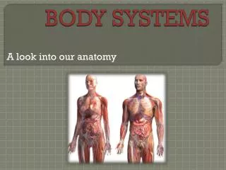
BODY SYSTEMS
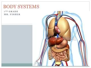
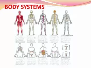
BODY SYSTEMS. BODY SYSTEMS. ORGAN SYSTEMS OF THE HUMAN BODY Nervous System – Receives, processes, & transmits information; coordinates all body systems. Endocrine System – Regulates homeostasis with chemicals known as hormones. Skeletal System – Supports and protects body parts.

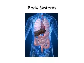
BODY SYSTEMS
E N D
Presentation Transcript
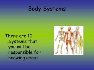
BODY SYSTEMS • ORGAN SYSTEMS OF THE HUMAN BODY • Nervous System – Receives, processes, & transmits information; coordinates all body systems. • Endocrine System – Regulates homeostasis with chemicals known as hormones. • Skeletal System – Supports and protects body parts. • Muscular System – Produces movement. • Integumentary System – Physical barrier against pathogens, injury, dehydration. • Circulatory System – Transports O2, CO2 , nutrients, wastes. • Respiratory System – Responsible for exchange of O2, CO2 • Immune System – Destroys pathogens • Digestive System– Breaks down food molecules to absorbable monomers • Urinary System – Washes blood; regulates blood volume • Reproductive System– Produces gametes; site of embryo development in females
INTEGUMENTARY SYSTEM Skin and its related structures—the hair, nails, and a variety of glands—make up the integumentary system. Pg. 933 Function of the Integumentary System • Serves as a gateway through which sensations such as pressure, heat, cold and pain are transmitted to the nervous system. • Serves as a barrier against infection and injury. • Regulates body temperature. • Removes wasteproducts • Protects against UV radiation.
INTEGUMENTARY SYSTEM 2 main layers 1. Epidermis • Composition and function of Outside layer: made up of dead cells containing keratin (tough fibrous protein) that waterproofs the skin; contains melanocytes (cells that produce melanin – pigment); protects against harmful UV rays • Composition and function of Inside layer: made up of living cells that produce keratin; replace outer layer dead cells 2. Dermis • Contains 7 structures: a.) collagen fibers b.) blood vessels c.) nerve endings d.) glands e.) sensory receptors f.) smooth muscles g.) hair follicles
INTEGUMENTARY SYSTEM 2 major types of glands: 1. Sweat glands: produce perspiration that removes water, salts and other compounds from the blood; cools body through evaporation 2. Sebaceous glands: produces oily secretion called sebum; helps keep skin flexible and waterproof Composition and function of Hypodermis: made up of fat and loose connective tissue; helps insulate the body Hair and nails Basic structure is keratin Function of hair: protects against UV rays, provides insulation, prevents dirt and other particles from entering the body Function of nails: protects the tips of fingers and toes Major Disease: Skin Cancer Cause of skin cancer: Excessive exposure to UV radiation Preventing skin cancer: wearing protective clothing and proper sun screen
INTEGUMENTARY SYSTEM Types of Burns
SKELETAL SYSTEM 1. Describe the function of the skeleton. Protect internal organs, provides for movement, stores mineral reserves, site for blood cell formation 2. What kind of system do bones and muscles provide in order to produce movement? System of levers 3. How many bones make up a normal human adult? 206 4. What are the 2 parts of the skeleton? What bones are found in each? Axial: skull, ribs, sternum, vertebral column Appendicular: clavicle, scapula, humerus, radius, ulna, carpals, metacarpals, phalanges, femur, patella fibula, tibia, tarsals, metatarsals 5. Describe the composition of bone. Calcium salts, Phosphorous, living cells and protein fibers
SKELETAL SYSTEM 6. What is periosteum? Tough layer of connective tissue What structure / substances are found within it? Blood vessels that carry oxygen and nutrients 7. Name 2 types of bone tissue. Compact and Spongy Where are they found within a bone? Compact bone is the outer layer. Spongy bone is the inner layer 8. What are Haversian canals? Tubes in compact bone that contain blood vessels and nerves What type of bone tissue contains them? Compact What structures pass through them? Blood vessels and nerves 9. Which type of bone tissue adds strength to bone without adding mass? Spongy bone 10. Describe each type of bone cell. Osteocyte: mature bone cells Osteoclast: cells that break down bone Osteroblast: cells that produce bone
SKELETAL SYSTEM 11. What is bone marrow? Soft tissue within bones What are the 2 types of bone marrow? Yellow and red Whatsubstances do each type of marrow produce? Yellow marrow produces fat cells. Red marrow produces red blood cells, white blood cells and cell fragments called platelets 12. What is cartilage? Connective tissue made up of tough collagen and flexible elastin How do embryonic skeletons and adult skeletons differ? Cartilage makes up almost the entire skeleton of the embryo, whereas in the adult it is mostly found in parts of the body that is flexible – ear, nose, rib cage 13. If cartilage does not contain blood vessels, how does it get nutrients? Through the diffusion of nutrients from tiny blood vessels in the surrounding tissue 14. How does cartilage support weight despite its extreme flexibility? Because it is dense and fibrous 15. What is ossification? The process of turning cartilage into bone When does it begin? Approximately 7 months before birth How areosteoblasts and osteocytes involved? Bone tissue forms as osteoblasts secrete mineral deposits that replace the cartilage in developing bones. When the osteoblasts become surrounded by bone tissue, they mature into osteocytes.
SKELETAL SYSTEM 16. Describe the growth process of long bones. Many long bones, including those of the arms and legs, have growth plates at either end. The growth of cartilage at these plates causes the bones to lengthen. Gradually, this new growth of cartilage is replaced by bone tissue, and the bones become larger and stronger. During late adolescence or early adulthood, the cartilage in the growth plates is replaced by bone, the bones become completely ossified, and the person “stops growing.” 17. Where is adult cartilage found? In adults, cartilage is found in those parts of the body that are flexible, such as the tip of the nose and the external ears. Cartilage also is found where the ribs are attached to the sternum, which allows the rib cage to move during breathing. 18. What is a joint? A place where one bone attaches to another bone Name the 3 classifications of joints. Immovable, Slightly movable and Freely movable Give an example of each. Immovable: bones of the skull Slightly moveable: vertebra and joints of the lower leg Freely moveable: Ball and socket, Hinge, Pivot and Saddle 19. What are ligaments? tough connective tissue which hold bones together in a joint Where are they found in the skeletal system? Attached to membranes that surround bones 20. What is the purpose of synovial fluid? enables the surfaces of the joint to slide over each other smoothly. What are bursae? small sacs of synovial fluid What substance is within one? Synovial fluid What is the purpose of a bursa? Reduces friction between the bones of a joints and act as tiny shock absorbers 21. Describe each of the following skeletal system disorders. Bursitis: Inflammation of a bursa Arthritis: inflammation of the joint Osteoporosis: loss of calcium that can lead to a weakening of the bones
SKELETAL SYSTEM • Joint • Ligament • Bursa • Cartilage • Tendons
Function • Movement • Regulate blood pressure • Move food through the digestive system • Power every movement in the body
Skeletal Muscle Characteristics • Attached to bones • Voluntary movements • Striated muscle- has alternating light & dark bands • Many nuclei • Long & slender • Complete skeletal muscle consists • Muscle fibers • Connective tissue • Blood vessels • nerves
Skeletal Muscle Characteristics • Muscle fibers are made up of bundles of cells • One muscle fiber cell is made of myofibril • Inside myofibril are 2 different filaments: • Actin (thin filaments) • Myosin (thick filaments) • The striations in muscle cells are formed by the Actin & Myosin
Skeletal muscles move bycontracting. Each muscle is composed of tightly packed bundles of muscle fibers, each containing proteins, which are responsible for muscle movement.Differentiation of muscles cells begins early inembryonic development. Myoblasts(A), the precursors of skeletal muscle cells, fuse together (B), to formmultinucleated cellswhich eventually develop intomyofibrils containingcontractile fibers ( C ).
Smooth Muscles • Involuntary movement • spindle shaped • has one nucleus
Found in … Eye Control the size of the pupil
Cardiac Muscle • Found only in the heart • Striated • Smaller than skeletal • One nucleus usually- some have 2 • Involuntary
How Muscles & Bones Interact • Tendons- tough connective tissue that joins muscle & bones together work like a lever and a fulcrum 2. Opposing muscle pairs- one muscle contracts, the other relaxes
Contraction of skeletal muscles helps move blood in veins toward the heart
Parts of the Circulatory System BLOOD HEART BLOOD VESSELS
FUNCTION OF THE CIRCULATORY SYSTEM • Transportation System of the Body • Oxygen • Carbon dioxide • Nutrients • Waste • Diffuse easily across cell membranes
2 Types of Circulatory Systems • Open Circulatory System = Invertebrates • Closed Circulatory System = Vertebrates
THE HEART • Made of cardiac muscle • 4 Chambers • 2 upper chambers • ATRIA • Receives blood • 2 lower chambers • VENTRICLES • Pumps blood
External Anatomy • Pericardium • a protective sac of tissue that surrounds the heart
Internal Anatomy • Myocardium • Thick layers of muscle that pump blood through the circulatory system. • Surrounded by 2 thin layers of epithelial and connective tissue
Internal Anatomy • Valves • Flaps of connective tissue between the atria and the ventricles. • When the ventricles contract, the valves close, which prevents blood from flowing back into the atria. • Blood moving from the atria holds the valves open.
Internal Anatomy • Septum • Divides the right side of the heart from the left side of the heart • Prevents the mixing of oxygen-poor and oxygen-rich blood L.A. SEPTUM R.A. R.V. R.V.
HEARTBEATThe Electrical Activity of the Heart 1 Sinoatrial node (Pacemaker) 2 Atrioventricular node 3 Atrioventricular Bundle 4 Left & Right Bundle branches 5 Bundle Branches
HEARTBEAT • Electrical activity initiates the heart muscle to contract • The pacemaker or sinuatrial node, which is situated in the upper wall of the right atrium initiates an impulse • The electrical impulse is picked up by a further electrical node called the atrioventricular node, which is situated in the lower part of the right atrium • The atrioventricular node picks up the impulse from the sinuatrial node and flows down the central wall of the heart (called the septum) • It is the passage of this electric conduction from the top of the heart over the atria through the septum and ventricles that causes the muscle to contract • It is the pattern of electrical conduction or electrical wave that is picked up on the electro-cardiogram or the ECG; the tracing of the heart's electrical activity.
Blood Vessels • Blood Vessels • Arteries, capillaries, and veins
ARTERIES • Large vessels that carry bloodfrom the heart to the tissues of the body • Superhighways of the circulatory system • Carry oxygen-rich blood, except for the pulmonary arteries • Thick walls that help them withstand the powerful pressure produced when the heart contracts and pushes blood into the arteries • Contain connective tissue, smooth muscle, and endothelium, allows an artery to expand under pressure • Contractions of the smooth muscle regulate the diameter of an artery
Aorta • First of a series of blood vessels that carry the blood on its round trip through the body and back to the heart • Blood leaves left ventricle and enters aorta • Loaded with oxygen from the lungs.
VEINS • Returns blood to the heart • Walls of veins contain connective tissue and smooth muscle. • Large veins contain valves that keep blood moving toward the heart. • Blood flow through the veins of the arms and legs often occurs against the force of gravity • If the walls around the veins weaken from lack of activity, the valves can weaken. This causes blood to pool in the veins, producing a condition known as varicose veins.
CAPILLARIES • Smallest of the blood vessels The walls of capillaries are only one cell thick, and most are so narrow that blood cells must pass through them in single file. Bring nutrients and oxygen to the tissues and absorbing carbon dioxide and other waste products from them
Circulation Through the Body • The heart functions as two separate pumps. 1. Pulmonary Circulation The right side of the heart pumps blood from the heart to the lungs. 2. Systemic Circulation The oxygen-rich blood then flows into the left side of the heart and is pumped to the rest of the body
Blood Pressure • When the heart contracts, it produces a wave of fluid pressure in the arteries. The force of the blood on the arteries' walls is known as blood pressure. • Medical workers can measure blood pressure with a device called a sphygmomanometer • A typical blood pressure reading for a healthy person is 120/80. • The first number is the systolic pressure—the force felt in the arteries when the ventricles contract. • The second number is the diastolic pressure—the force of the blood felt in the arteries when the ventricles relax.