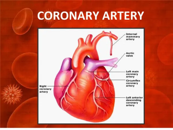
Basic Coronary Artery Anatomy
Medtronic Fellows PCI Primer. Basic Coronary Artery Anatomy. Paul Fefer, MD. Interventional Cardiology Unit Sheba Medical Center, Tel Hashomer Courtesy of Frederick Feit, MD. The Cardiovascular Research Foundation. Transcatheter Cardiovascular Therapeutics. Basic Coronary Artery Anatomy.

Basic Coronary Artery Anatomy
E N D
Presentation Transcript
Medtronic Fellows PCI Primer Basic Coronary Artery Anatomy Paul Fefer, MD. Interventional Cardiology Unit Sheba Medical Center, Tel Hashomer Courtesy of Frederick Feit, MD The Cardiovascular Research Foundation Transcatheter Cardiovascular Therapeutics
Basic Coronary Artery Anatomy Sternocostal Aspect
Basic Coronary Artery Anatomy Diaphragmatic Aspect
Right Coronary Artery Basic Anatomy • OriginRight aortic sinus (lower origin than LCA) • CourseDown right AV groove toward crux of the heart, gives off PDA (85%) from which septals arise, continues in LAV groove giving off posterior LV branches (posterolaterals). PDA may originate more proximally, bifurcate early or be small with part of “its territory” supplied by an acute marginal branch. • Supplies25% to 35% of Left Ventricle
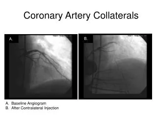
Right Coronary Artery Other Branches • Conus Arteryusually very proximal; (~50% have a separate origin)-courses anteriorly and upward over the RV outflow tract toward the LAD. May be an important source of collaterals. • SA Nodal Artery(~60%) usually 2nd branch of RCA-courses obliquely backward through upper portion of atrial septum and anteromedial wall of the RA-supplies SA node, usually RA and sometimes LA.
Right Coronary Artery Other Branches • Right Ventricular (Acute Marginal) Branches)Arise from mid RCA; supply anterior RV; may be a collateral source. • AV Nodal ArteryArises at or near crux; supplies AV node. • PDASupplies inferior wall, ventricular septum, posteromedial papillary muscle.
Right Coronary Artery Optimal View(s) • LAO (30) Cranial(30)particularly for distal bifurcation (AP Cranial may be better). • RAOmain shaft; cranial enhances distal vessels and very proximal; caudal may help with Shepherd’s crook. • Lateralbifurcations with RV branches-distal bifurcation, particularly with cranial.
LAO Cranial Angiogram of RCA PDA Acute Marginal
Main RCA Acute Marginal Native RCA Lateral View Demonstrating Origin of Acute Marginal
AP Cranial View of Distal RCA RPL 2 AV Groove RPL 1 PDA
Left Coronary Artery Left Main Coronary Artery • Originupper portion of left aortic sinus just below the sinotubular ridge. Typically 0-10 mm in length. Rarely no LM (separate origins). • Catheterization Technique“The Judkins’ 4-Left coronary catheter will find the LCA orifice unless thwarted by the operator”. Just in case-other Judkins sizes for smaller or larger aortas; Amplatz, XB type curves. Watch for “damping”; For separate ostia-separate catheters, larger for Cx, or counterclockwise rotation for LAD. • Optimal ViewsLAO caudal and cranial; AP-caudal, cranial or flat. Limit views. May need IVUS
7th Annual Interventional Cardiology Self-Assessment Course at TCT2004 Basic Coronary Artery Anatomy: Frederick Feit, M.D. Sternocostal Aspect
7th Annual Interventional Cardiology Self-Assessment Course at TCT2004 Basic Coronary Artery Anatomy: Frederick Feit, M.D. Diaphragmatic Aspect
Left Anterior Descending Artery • Coursedown the anterior interventricular groove-usually reaches apex. In 22% of cases does not reach apex. • Branchesseptals and diagonals-supply lateral wall of LV, anterolateral papillary muscle; 37% have median ramus (courses like 1st diagonal). • LADSupplies anterolateral, apex and septum; ~45%-55% of left ventricle.
Left Circumflex Artery • Originfrom distal LMCA. • Coursedown distal left AV groove. • Branchesobtuse marginal, posterolaterals-supply posterolateral LV, anterolateral papillary muscle. SA node artery-38%. • Supplies15%-25% of LV, unless dominant (supplies 40-50% of LV).
Left Coronary Artery Optimal Views • AP (30)CaudalLMCA, proximal LAD, Cx, distal LAD. Poor for mid LAD- RAO may be useful. • AP (40)CranialLMCA, LAD, diagonals, septals, distal Cx-may need RAO to separate LAD and Cx. • (45)LAO (35) CranialLMCA, LAD, diagonals, septals, and distal Cx. • (45)LAO (30) CaudalLMCA, Cx,and prox LAD. • Laterals (cranial, caudal)may be helpful.
AP Caudal View of LCA LAD Circ
AP Cranial View of LCA Cx LAD Septal Diagonal
AP Cranial LCA Angiogram LMCA Cx Diagonals Septal LAD
LAO Cranial View of LCA Circ LAD Diagonal
LAO Caudal View of LCA Median Ramus LAD Circ
Dominance: • Definition 1:the coronary artery which reaches the crux of the heart and then gives off the PDA • Definition 2: (Allows for codominance)the artery which gives off the PDA as well as a large posterolateral branch
7th Annual Interventional Cardiology Self-Assessment Course at TCT2004 Basic Coronary Artery Anatomy: Frederick Feit, M.D. LeftDominantCirculation
LCA Angiogram Dominant Cx AP Caudal Prox LAD LM Occluded Median Ramus Distal LAD OM Distal Cx
LCA Angiogram-Dominant Cx LAO-Caudal Prox LAD OccludedMedianRamus LM Prox Cx Distal LAD LPDA
The Coronary Arteries Are Complementary • Large PDA Small LAD • Huge Cx (posterolaterals) Small RCA continuation in AV Groove • Etc, etc, etc…..
BYPASS GRAFTS • SVGLeft coronary grafts generally arise from left side of the aorta. Best cannulated with Judkins’ Right, IMA, LCB or MP. • Right sided grafts-arise from right side of the aorta-MP usually best. • IMAdon’t forget to check subclavians. All distal vessels must be accounted for; op notes and old films are extremely helpful.
SVG-OM-LAO Caudal Demonstrating Graft Ostium Ostium
SVG-OM 1 AP Caudal Demonstrating Anastomosis SVG
LIMA to LAD Origin from left subclavian (AP Cranial)
LIMA to LAD Distal Anastomosis-AP Cranial LIMA LAD
Native LCA AP Caudal Stump of original SVG to OM 1
SVG to OM Lesion 2 Lesion 1
SVG to OM Slight change of view to demonstrate unequivocal severity of lesion Lesion 2 Lesion 1
SVG to RCA Multipurpose Technique -LAO SVG
LCA AP Caudal Severe stenosis Distal LADwith slow flow
Thrombus In LAD Post-NTG-Thrombus has migrated distally but still adherent Thrombus
AP Cranial Thrombus In LAD Thrombus
Calcified Native RCA (LAO Cranial)“Bone Island” Simulating Thrombus
Myocardial Bridging Intramyocardial Segment • Almost always LAD • Occurs in 5-12% of patients • Usually not hemodynamically significant
Myocardial Bridging LCA-RAO Projection LAD Diastole LAD Systole