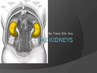
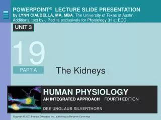
The Kidneys
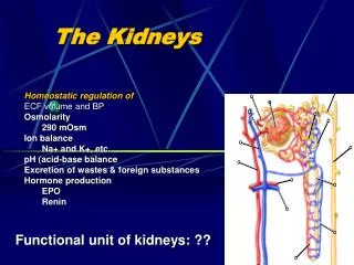
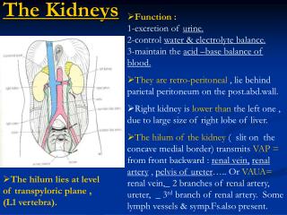
The Kidneys. 19. Functions of the Kidneys. Regulation of extracellular fluid volume and blood pressure - works with CV system to ensure tissues get enough oxygen and BP is within normal values Regulation of osmolarity – blood osmolarity needs to be maintained around 290mOsM

The Kidneys
E N D
Presentation Transcript
The Kidneys 19
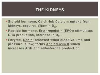
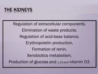
Functions of the Kidneys • Regulation of extracellular fluid volume and blood pressure - works with CV system to ensure tissues get enough oxygen and BP is within normal values • Regulation of osmolarity – blood osmolarity needs to be maintained around 290mOsM • Maintenance of ion balance - in response to diet urinary loss helps to maintain proper levels of Na+, K+, Ca 2+ . • Homeostatic regulation of pH – they remove either H+ or HCO3- as needed, they don’t correct pH imbalances as effectively as the lungs • Excretion of wastes – removes waste molecules dissolved in the plasma like urea (from amino acid breakdown), uric acid (nucleic acid turnover), and creatine (from creatine phosphate breakdown). • Production of hormones – erythropoietin (signal RBC production), renin (influence BP and BV), and vitamin D conversion to control Ca 2+ .
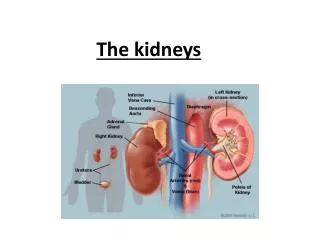
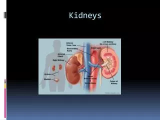
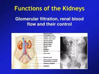
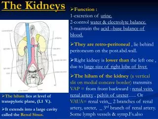
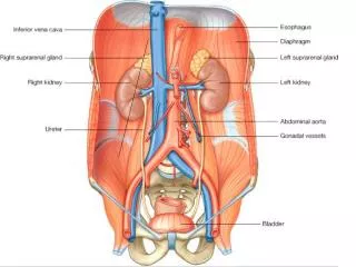
Anatomy: The Urinary System Figure 19-1a
Anatomy: The Urinary System Cortico & juxtamedullary nephrons Figure 19-1c
Anatomy: The Urinary System Figure 19-1d–e
Anatomy: The Urinary System Figure 19-1g–h
Anatomy: The Urinary System Figure 19-1f
Kidney Function Peritubular capillaries Distal tubule Efferent arteriole Glomerulus F Afferent arteriole Bowman’s capsule Proximal tubule Loop of Henle KEY = Filtration: blood to lumen F To renal vein Collecting duct Figure 19-2 (1 of 4)
Kidney Function Peritubular capillaries Distal tubule Efferent arteriole R Glomerulus F R Afferent arteriole Proximal tubule Bowman’s capsule R R R Loop of Henle KEY F = Filtration: blood to lumen To renal vein = Reabsorption: lumen to blood R Collecting duct Figure 19-2 (2 of 4)
Kidney Function Peritubular capillaries Distal tubule Efferent arteriole S R Glomerulus F R S Afferent arteriole Proximal tubule Bowman’s capsule R S R R Loop of Henle KEY F = Filtration: blood to lumen To renal vein = Reabsorption: lumen to blood R Collecting duct = Secretion: blood to lumen S Figure 19-2 (3 of 4)
Kidney Function Peritubular capillaries Distal tubule Efferent arteriole S R Glomerulus F R S Afferent arteriole Proximal tubule Bowman’s capsule R S R R Loop of Henle KEY F = Filtration: blood to lumen To renal vein = Reabsorption: lumen to blood R Collecting duct = Secretion: blood to lumen S E = Excretion: lumen to external environment E To bladder and external environment Figure 19-2 (4 of 4)
Kidney Function The urinary excretion of substance depends on its filtration, reabsorption, and secretion Figure 19-3
Filtration Fraction Figure 19-5
Filtration at the glomerulus • Podocytes wrap around fenestrated capilaries creating filtration slits at the glomerulus.
Forces that Influence Filtration • Hydrostatic pressure (blood pressure) – pressure of flowing blood in glomerular capillaries is 55mmHg, it favors the movement of filtrate into Bowman’ Capsule • Colloid osmotic pressure –Plasma proteins that enter the capsule create a gradient the favors movement back into the capillaries • Fluid pressure created by fluid in Bowman’s capsule – The fluid build-up in the enclosed capsule creates a gradient that favors movement back into the capillaries The combination of these factors causes filtration to return plasma into the capillaries and allow for only 20% of the filtered plasma to move along the tubules.
Filtration Filtration pressure in the renal corpuscle depends on hydrostatic pressure, colloid osmotic pressure, and fluid pressure Figure 19-6
Filtration Autoregulation of glomerular filtration rate takes place over a wide range of blood pressure Figure 19-7
Glomerular Filtration Rate Changes GFR is controlled by a • myogenic response, • tubuloglomerular feedback, • hormones and autonomic neurons • Changing resistance in arterioles altes the filtration coefficient
Juxtaglomerular Apparatus Juxtaglomerular cells and Macula densa monitor blood flow and blood pressure along the arteioles. They send chemical signals needed to restore the proper filtration rate Figure 19-9
Tubuloglomerular Feedback Distal tubule Efferent arteriole Glomerulus Bowman’s capsule GFR increases. 1 Proximal tubule Macula densa 2 Flow through tubule increases. 1 Afferent arteriole Granular cells 2 2 Collecting duct Loop of Henle Figure 19-10, steps 1–2
Tubuloglomerular Feedback Distal tubule Efferent arteriole Glomerulus Bowman’s capsule GFR increases. 1 Proximal tubule Macula densa 2 Flow through tubule increases. 4 1 3 Flow past macula densa increases. Afferent arteriole Granular cells 3 2 2 4 Paracrine diffuses from macula densa to afferent arteriole. Collecting duct Loop of Henle Figure 19-10, steps 1–4
Tubuloglomerular Feedback Distal tubule Efferent arteriole Glomerulus Bowman’s capsule GFR increases. 1 Proximal tubule Macula densa 2 Flow through tubule increases. 4 1 5 3 Flow past macula densa increases. Afferent arteriole Granular cells 3 2 2 4 Paracrine diffuses from macula densa to afferent arteriole. 5 Afferent arteriole constricts. Resistance in afferent arteriole increases. Collecting duct Loop of Henle Figure 19-10, steps 1–5 (2 of 4)
Tubuloglomerular Feedback Distal tubule Efferent arteriole Glomerulus Bowman’s capsule GFR increases. 1 Proximal tubule Macula densa 2 Flow through tubule increases. 4 1 5 3 Flow past macula densa increases. Afferent arteriole Granular cells 3 2 2 4 Paracrine diffuses from macula densa to afferent arteriole. 5 Afferent arteriole constricts. Resistance in afferent arteriole increases. Collecting duct Hydrostatic pressure in glomerulus decreases. Loop of Henle GFR decreases. Figure 19-10, steps 1–5 (4 of 4)
Reabsorption 1 Na+ is reabsorbed by active transport. Filtrate is similar to interstitial fluid. 2 Electrochemical gradient drives anion reabsorption. Na+ 1 2 Anions 3 Water moves by osmosis, following solute reabsorption. 3 H2O Concentrations of other solutes increase as fluid volume in lumen decreases. Permeable solutes are reabsorbed by diffusion. 4 4 K+, Ca2+, urea Tubular epithelium Extracellular fluid Tubule lumen Principles governing the tubular reabsorption of solutes and water. Sodium and water always follow each other.Transepithelial transport-(passing through cells)-Substances cross both apical and basolateral membraneParacellular pathway(passing around cells)-Substances pass through the junction between two adjacent cells Figure 19-11
Reabsorption Saturation of mediated transport Transport rate is proportional to plasma concentration until transport saturation=renal threshold Figure 19-14
Reabsorption Glucose handling by the nephron This graph does not show saturation at Bowman’s capsule Figure 19-15a
Reabsorption Saturation is reached within the proximal tubule Figure 19-15b
Reabsorption Excretion rate shows that no glucose is excreted with when plasma glucose concentration is low. Figure 19-15c
Reabsorption Glucose is not secreted When filtration and reabsoption are equal and below threshold there is no secretion. Above that results in glucosuria or glycosuria Figure 19-15d
Secretion • Transfer of molecules from extracellular fluid into lumen of the nephron - dependent on membrane transport proteins to move organic compounds • Active process – move against concentration gradient and use secondary active transport to move into lumen • Secretion of K+ and H+ is important in homeostatic regulation • Enables the nephron to enhance excretion of a substance – adds to the substances collected during filtration, making excretion more effective • Competition decreases penicillin secretion – doctors combined probenecid with penicillin so it would compete for the transporter protein and keep the kidneys from clearing penicillin so quickly.
Excretion • Excretion = filtration – reabsorption + secretion • Clearance • Rate at which a solute disappears from the body by excretion or by metabolism • Non-invasive way to measure GFR • Inulin and creatinine used to measure GFR
Inulin Clearance Efferent arteriole Filtration (100 mL/min) Glomerulus Peritubular capillaries 2 Afferent arteriole 1 Nephron Inulin molecules KEY = 100 mL of plasma or filtrate 3 100 mL, 0% inulin reabsorbed 1 Inulin concentration is 4/100 mL 2 GFR = 100 mL /min 3 100 mL plasma is reabsorbed. No inulin is reabsorbed. 4 Inulin clearance = 100 mL/min 4 100% of inulin is excreted so inulin clearance = 100 mL/min 100% inulin excreted Inulin=polysaccharide; 100% of it is excreted so it is used to measure glomerular filtration rate Clearance is the rate at which a solute disappears from the body Figure 19-16
Excretion The relationship between clearance and excretion is that clearance is the rate of excretion. Different substance have difference clearance.
Micturition The storage of urine and the micturition reflex Figure 19-18a
Micturition 1 2 3 Stretch receptors fire. Parasympathetic neurons fire. Motor neurons stop firing. Smooth muscle contracts. Internal sphincter passively pulled open. External sphincter relaxes. (b) Micturition Higher CNS input may facilitate or inhibit reflex. Stretch receptors Sensory neuron 1 Parasympathetic neuron 2 3 + – Motor neuron Tonic discharge inhibited Internal sphincter 2 3 External sphincter Figure 19-18b