Download
1 / 39
430 likes | 878 Vues
Structures and Related Common Pathologies of the Knee. Functional Requirements of the Knee. Stability and The knee must be stable enough to withstand high loads Walking on level surfaces = Stair climbing = The knee must transmit loads from the upper body and thigh to the lower leg

E N D
Functional Requirements of the Knee • Stability and • The knee must be stable enough to withstand high loads • Walking on level surfaces = • Stair climbing = • The knee must transmit loads from the upper body and thigh to the lower leg • Mobile enough to permit wide range of ROM for ADL’s • Walking = 0-65 • Climbing stairs 0-83 • Squatting to lift object = 0-115
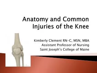
Anatomy of the Knee • The knee joint is called the tibiofemoral joint • Largest joint in the body • Tibiofemoral joint typically has • Comprised of the femur and tibia that are connected by four strong ligaments that limit motion in any direction • Synovial joint
Anatomy of the Knee • Femoral condyles rest on top of tibial surface to form • The patella glides through a special groove formed by the two femoral condyles called the
Menisci • Menisci are found on the • Medial and lateral menisci e join • Horseshoe-shaped pieces of cartilage which increase joint concavity and contact surface • Menisci spread forces out across the joint, transmitting some of the load to the articular cartilage of the tibial plateau • Combination of menisci and articular cartilage of the tibial plateu produce a nearly frictionless gliding surface • The menisci aid in lubrication, nutrition, and shock absorption (decrease joint stress)
Patellar Anatomy • The patella, is embedded in the tendon of quadriceps femoris, and is located anterior to the tibio-femoral articulation
Patella Function • The patella is part of the knee joint extensor mechanism and works as a lever to provide a greater mechanical advantage for the muscles that extend the knee
Muscles, Nerves, and Blood Vessels • The Extensor Mechanism: • Patella, patellar tendon, quadriceps tendon, and quadriceps muscles • Popliteal Nerve: • Located in back of the knee and travels to the lower leg and foot, supplying sensation and muscle control. • The Popliteal Artery (carries blood to the leg and foot) and Popliteal Vein (carries blood back to the heart). • If the popliteal artery is damaged beyond repair, it is very likely the leg will not be able to survive.
Knee Stability • The ligaments, joint capsule and menisci are the “static” stabilizers of the knee • The major ligaments of the knee are: • Anterior Cruciate Ligament (ACL) • Posterior Cruciate Ligament (PCL) • Medial Cruciate Ligament (MCL) • Lateral Collateral Ligament (LCL)
Anterior Cruciate Ligament • Attaches on the anterior portion of the tibia in a superior-posterior direction and attaches on the posterior lateral femoral condyle • Acts to limit knee rotation • Resists anterior displacement of the tibia on the femur • Keeps the tibia
Posterior Cruciate Ligament • It attaches from the posterior portion of the tibia to the medial condyle of the femur • Resists posterior displacement of the tibia on the femur • Keeps the tibia from
Anterior and Posterior Cruciate Ligaments • Named cruciate ligaments because they cross each other (like the limbs of the letter X). Front of right knee
Medial Collateral Ligament • The MCL attaches from the medial condyle of the femur to the medial condyle of the tibia • Works to resist and external rotation forces
Lateral Collateral Ligament • Attaches from the lateral condyle of the femur to the fibula • Works to resist
Mechanism of ACL Injury • Most people who rupture their ACL can recall the exact moment at which they felt it pop: changing direction, pivoting or cutting in sports like soccer, landing from a jump in sports such as basketball, or falling while skiing • The 4 "classic" symptoms reported with an ACL tear are: • A "pop" from inside the knee • Knee “giving-away” at the time of injury • Knee swells immediately, or within 12 hours • Hemarthrosis: bleeding in the joint
Mechanism of ACL injury • With injury, the tibia slides anteriorly on the femur, causing the knee to "give-way". Most lax at 30, but injuries can occur from 15 -45 • When the ACL is ruptured, there is usually damage to other knee structures as well: • Medial or lateral collateral ligaments • Menisci • Women are much more susceptible to ACL injuries than men (Q-Angle)
Quadriceps Angle or Q-Angle • Formed by a line drawn from the ASIS to the center of the kneecap, and a line drawn from the center of the knee cap to the Tibial tuberosity. • Normal: Males=10° Females=15
Rehabilitation for Cruciate Ligament Injuries • Takes about six months for graft to heal • Hamstring or Patellar Tendon • Focus on closed-chained exercises • Foot is against a surface • Squats, step-ups, lunges • Initially avoid rotational exercises • Priority on re-establishing proprioception
Mechanism of PCL Injury • The PCL is susceptible to rupture in the face of any severe, direct blow to the front of the knee while the knee is flexed (such as from "dashboard knee" in a car accident) • The incidence of posterior cruciate ligament injuries is far less common than that of anterior cruciate ligament injuries.
Mechanism of MCL Injury • Blow to the outside of the knee • Severe outward twist • A complete rupture, will usually involve the ACL
Mechanism of LCL Injury • Blow to the inside of the knee • Severe rotational or direct force • Injuries to the LCL are must less prevalent than those to the MCL • A complete rupture will usually involve the ACL, PCL, Menisci, IT band and Biceps Femoris
Rehabilitation for Collateral Ligament Injuries • Takes about six months for graft to heal • Hamstring or Patellar Tendon • Focus on closed-chained exercises • Distal joint is fixed • Squats, step-ups, lunges • Avoid rotational exercises until late in program • Priority on re-establishing proprioception
Mechanism of Meniscal Injuries • Meniscal tears and issues are about the most common traumatic lesions of the knee • The usual mechanism of injury is a twisting force on a weight bearing flexed knee. • May be traumatic or degenerative with a mild injury producing the problem. • Menisci penetrated by nerves, but almost no blood vessels • When menisci are torn, there is pain but no bleeding • Torn meniscus does not heal
Rehabilitation for Meniscal Injuries • Focus on re-gaining muscular strength and neuromuscular control • Return to activity is relatively quick • Partial menisectomy (six to 14 days) • Repaired meniscus is a little longer (about two months)
Patellofemoral Pain Syndrome (PFPS) • Also known as Chondromalacia Patella • Softening and deterioration of the articular cartilage on the back of the patella • May be swelling around the kneecap and a grating sensation when flexing or extending the knee • Characterized by pain in the anterior aspect of the knee, aggravated by knee extension activities such as ascending/descending stairs, squatting, rising from a chair, jumping • May affect one or both knees • Highest incidence is in females • Incidence among athletes >25%.
Contributing Factors to Patellofemoral Pain Syndrome • Q-angle • Patella Alta/Baja • Hip Muscle Weakness • Tight IT band • Femoral Adduction and Medial rotation • Overuse/Overload • Vastus Medialis Weakness or imbalances between Vastus Medialis and Vastus Lateralis • Subluxation/Dislocation
Patella Alta/Baja • Patella Alta: • Patella positioned higher and creates a greater chance that quads can dislocate the patella • Patella Baja: • Patella positioned lower & tends to lead to ’d compressive forces.
Hip Muscle Weakness • Weak musculature or poor neuromuscular control increased medial rotation and adduction of the hip dynamic activities • Poor motor control may cause changes in pelvic and femoral position altered contact pressure at the knee. • Decreased muscle endurance may contribute to poor motor control at the hip. • In theory, poor proprioception may impact positioning of the femur
Patellofemoral Pain Syndrome Treatment Strategies • Closed chain exercises in pain-free range of motion to strengthening quadriceps/hamstrings • Need to re-establish correct patellar tracking • Stretching
Patellofemoral Pain Syndrome Treatment Strategies Hip Abduction & External Rotation
Patellar Injuries and Other Issues • Patellar Tendon Rupture
Patellar Injuries and Other Issues • Osgood-Schlatter • Avulsion fracture at the attachment of the patellar tendon at the tibial tubercule
Patellar Injuries and Other Issues • Patellar Subluxation/Dislocation
Patellar Injuries and Other Issues • Patellar Fracture
Knee Replacement Rehabilitation • Focus on re-gaining muscular strength, range of motion and neuromuscular control (proprioception) • Exercises • Primarily closed chain • Squats, lunges, step-ups, leg press • Proprioception • Bosu, wobble boards, single leg
Knee Stability • The muscles about the knee are important “dynamic” stabilizers. • For optimal knee function, support must come from both the integrity of the static stabilizers & good strength of the dynamic stabilizers • Important mechanoreceptors may get disrupted with injury or surgery • Specialized nerve endings that respond to mechanical pressure or deformation and relay that information centrally in the nervous system • During and after the rehab process, improving knee proprioception and neuromuscular control is imperative.