Download
1 / 12
130 likes | 524 Vues
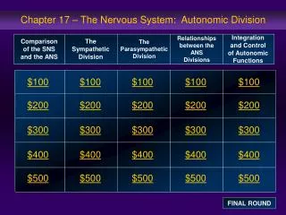
Ch 17: Autonomic Division of NS. Compare and contrast the structures of the sympathetic and the parasympathetic divisions, including functions and neurotransmitters. Show the levels of integration in the ANS, and compare these with the SNS. Overview of ANS. Pathway for Visceral Motor Output

E N D
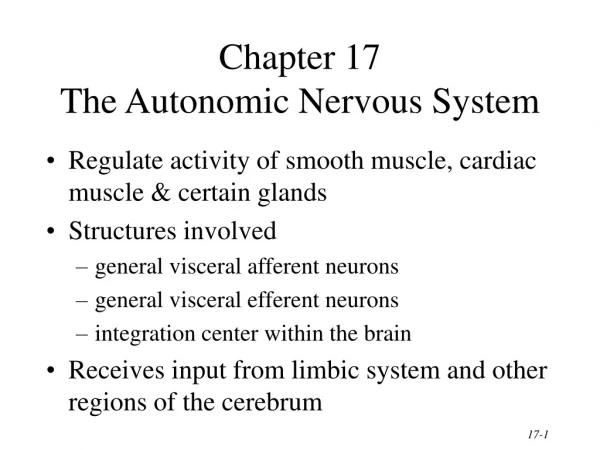
Ch 17: Autonomic Division of NS Compare and contrast the structures of the sympathetic and the parasympathetic divisions, including functions and neurotransmitters. Show the levels of integration in the ANS, and compare these with the SNS.
Overview of ANS Pathway for Visceral Motor Output ANS has two antagonistic divisions: • Sympathetic • Parasympathetic ANS output always involves two neurons between spinal cord (CNS) and effector.
Synapsing takes place in ganglia Naming of neurons: ? Fig 17.3
Sympathetic Division Thoracolumbar division Preganglionic neurons (cell bodies) located between T1 & L2 of spinal cord Ganglionic neurons (cell bodies) in ganglia near vertebral column Paravertebral ganglia = sympathetic chain ganglia Prevertebral ganglia = collateral ganglia Special case: adrenal medulla Effects of Sympathetic Division?
Special Case:Adrenal medulla Fig. 17-6 Modified sympathetic ganglion Terminus for neuron #1, stimulates specialized 2nd order neurons with very short axons in adrenal medulla to release NT into blood stream (= hormones) Epinephrine (adrenalin) ~ 80% and norepinephrine (noradrenalin) Endocrine effects are longer lasting than nervous system effects
Sympathetic Neuroeffector Junctions Differ from somatic neuromuscular junctions Varicosities Fig 17-6
Summary of Sympathetic Division A. Neuron #1 is short, neuron #2 is long B. Synapsing occurs in prevertebral chain ganglia or paravertebral collateral ganglia C. Neuron #1 releases Ach, usually neuron #2 releases NE D. Prepares for emergency action, excitatory to many organs, inhibitory to others ( digestive for example) E. Effects very widespread and somewhat persistent
Para – SympatheticDivision Craniosacral division Preganglionic neurons (cell bodies) located in brain stem & sacral segments of spinal cord Ganglionic neurons (cell bodies) in ganglia near target organs: Intramural ganglia Effects of parasympathetic division ?
Summary of Parasympathetic Division A. Neurons #1 are long, come from the brain stem or sacral spinal cord, run with the spinal or pelvic nerves and produce ACh. B. Neurons #2 are short, produce ACh, and may be either excitory or inhibitory.
Anatomy of Dual Innervation Each organ receives innervation from sympathetic and parasympathetic fibers Fibers of both divisions meet & commingle at plexuses (fig 17-9) to innervate organs close to those centers Names of plexuses derived from locations or organs involved