Effective Techniques for Post-Mortem Joint Fluid Collection in Pigs
380 likes | 463 Vues
Learn detailed steps for aseptically collecting joint fluid from stifle and hock areas in pigs post-mortem to ensure accurate diagnostics. Proper cotton rope usage for oral fluid collection is also discussed.

Effective Techniques for Post-Mortem Joint Fluid Collection in Pigs
E N D
Presentation Transcript
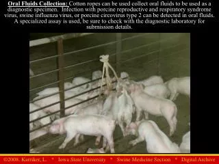
Oral Fluids Collection: Cotton ropes can be used collect oral fluids to be used as a diagnostic specimen. Infection with porcine reproductive and respiratory syndrome virus, swine influenza virus, or porcine circovirus type 2 can be detected in oral fluids. A specialized assay is used, be sure to check with the diagnostic laboratory for submission details.
Euthanasia: Proper Captive Bolt Gun Placement Draw a line from medial canthus of the eye to the medial side of opposite ear, repeat on other side. Where the lines cross is the proper place (yellow circle) to place the bolt gun. Be sure to have the end of the gun flat against the skull so that the barrel is perpendicular to the surface of the skull. Proper placement should penetrate the brain and brainstem. Ensure proper cartridge size
Captive bolt gun: Utilizes gun powder charges to a drive bolt through the brainstem for euthanasia. 130 milligrain charge adequate for most pigs Traditional style captive bolt gun
Post-mortem sample collection technique for aseptically collecting joint fluid from both the stifle and hock Step 1: Make a slit through the skin along midline (not directly over the joints) and skin the leg all the way down past the hock
Post-mortem sample collection technique for aseptically collecting joint fluid from both the stifle and hock Step 2, Stifle Samples: Once skinned, locate the patella and then make three deep slices with your knife around the stifle It is important to make your cuts all the way to the underlying bone Patella
Post-mortem sample collection technique for aseptically collecting joint fluid from both the stifle and hock Step 2 Images Palpating the patella Finishing making the 3rd cut
Post-mortem sample collection technique for aseptically collecting joint fluid from both the stifle and hock Step 3: Along the medial cut make sure you have sliced down through all tissue layers to the bone Locate the patella and slide it laterally, thus exposing the stifle joint without contaminating it. Patella pulled laterally with muscle Open stifle joint (note the presence of the patellar grove)
Post-mortem sample collection technique for aseptically collecting joint fluid from both the stifle and hock Step 4: Collect sample from the joint by using a sterile syringe and a 20 g x 1” needle
Post-mortem sample collection technique for aseptically collecting joint fluid from both the stifle and hock Step 5: Make a tiny incision in the joint capsule to allow access for a swab. Don’t stab into the joint. Use a new clean knife or sterile scalpel for this incision. Use a sterile swab to obtain a sample for culture.
Post-mortem sample collection technique for aseptically collecting joint fluid from both the stifle and hock Step 6 hock samples: Use the most distal cut that was originally made for the stifle sample to access the hock joint Patella
Post-mortem sample collection technique for aseptically collecting joint fluid from both the stifle and hock Step 7: Grab the belly of the transected muscle and pull it distally, taking your knife and cutting connective tissue as you pull, exposing the hock joint
Post-mortem sample collection technique for aseptically collecting joint fluid from both the stifle and hock Step 8: At this point you should be able to visualize the hock joint and collect samples without contaminating the joint.
Post-mortem sample collection technique for aseptically collecting joint fluid from both the stifle and hock Transfer samples to appropriate packaging and label with pig identifier.
This is a common method for restraining and bleeding pigs that are too small for the snare method.
Vaccination schedule at a sow farms: Help to keep pre-farrowing vaccinations and feed adjustments on schedule even when employees most familiar with the process are off for the day.
Posted treatment protocols: These are critical to getting consistent results and proper treatment over time. Without a consistent approach to treatment, response can not be evaluated and refinements are tough to make. Compounding is illegal: Mixtures often mask an underlying problem or risk factor that needs to be corrected.
The blue device is checking for the amount of deterioration that has occurred to the nasal turbinates of the pig. The deeper the device is able to enter the turbinates, the greater degree of damage or loss of turbinate function has occurred.
Needle-free injection system: when pressed firmly against the hide of an animal, automatically propels, via a pressurized system, a dose of treatment through the skin and into muscle tissue. Injector Carbon-dioxide canister attaches here Pressure gauge
Full user harness is beneficial when moving through open pens or when injecting multiple crated animals Needleless Injector System System without harness is beneficial when working in a stationary location or farrowing room handling piglets Pressure Amplifier CO2 Cylinder
Needleless injector used for iron dextran injections in pre-weaning piglets Note: All injections are only given in the neck muscle of the piglet
Blood collection on mature swine Hog snare is placed around the snout and behind the incisors to restrain the animal Vacutainer® blood collection system (similar to human collection system)
Blood collection via ear swabbing The blood collected on the swab can be stored in a falcon tube containing sterile saline and then submitted to a diagnostic lab. Samples collected in this manner have substantial dx limitations. Mainly used for daily monitoring of boars during semen collection Collecting blood on a sterile, synthetic swab after puncturing the ear vein.
Proper location for administering TB antigen when testing swine
Back Fat Determination Using Real-time Ultrasound Technology Probe is placed at the 10th or last rib of the animal. The visual image of the underlying fat and muscle is saved and measured to determine carcass traits of the animal
Swine carcass incinerator: Diesel-fuel powered machine that is used to reduce animal carcasses to ash. Use of incinerators reduce site traffic and are thus more ideal for biosecurity reasons.
Bedding, usually sawdust, is used for increased traction during transfer Portable Swine Transfer Chute: Used to transfer swine directly between two semi trailers.
Support boards to allow human access to transferring swine without entering either vehicle. Portable Swine Transfer Chute: Used to transfer swine directly between two semi trailers.
Trailer dryer:Drying is used to reduce the pathogen load present on the trailer after washing and disinfection.
Truck Wash Facility This trailer completed wash cycle, parked on clean side, and on incline to drain water Trailer wash bays Dirty trailers wait and enter wash bay from this side only
Rendering Vehicle:This vehicle has recently emptied a disposal bin without transferring the contents completely and effectively into the holding compartment on the truck. This creates a biosecurity concern, a safety risk for the driver and a public relations problem.
Farm cats and pigs don’t mix. Cats are not effective rodent control and can spread disease to the pigs on the site
Bones from a deceased pig. The dead animal was not located in the pen of pigs and was consumed. This is illegal in most states, a biosecurity risk, and poor welfare.
Temporary swine identifiers/markers; examples of products that are commonly used to identify pigs during treatment, handling, etc. Such marking devices can be found in several forms.
Ear notch identification system:a commonly used practice in purebred, replacement stock or small producer herds, that involves cutting notches in piglet ears at an early age 3+3+1+81=88 1+1+3=5 Right designates litter number Left designates pig number
Animals marked to indicate treatments or vaccinations have been administered