Download

1 / 73
730 likes | 983 Vues
Lower Extremity Wounds: The role of the vascular technologist. Jesse Thomas, RVT UNC Health Care. Disclosures No relevant conflicts of interest to declare. Objectives. Review types of wounds Discuss risk factors Role of Duplex Imaging Role as a Technologist

E N D
Lower Extremity Wounds: The role of the vascular technologist Jesse Thomas, RVT UNC Health Care
Objectives • Review types of wounds • Discuss risk factors • Role of Duplex Imaging • Role as a Technologist • This presentation will NOT address the use of ultrasound as a wound management and/or treatment tool.
Types of Wounds • Arterial • Venous • Neuropathic • Small vessel/Vasculitis • Pressure ulcers
Arterial • Ischemic wounds • Result of inadequate blood supply • Tissue hypoxia and tissue damage • Most commonly result of atherosclerotic disease (PAD)
PAD • Narrowing of arteries to the limbs that reduces blood flow • More common in LE • Atherosclerosis – build up of fatty deposits (plaque)
Arterial • Risk Factors • High cholesterol • Aging • HTN • Diabetes • Smoking • Family hx of cardiovascular disease • Obesity
PAD • Approximately 8 million people in the US • 12-20% in those >60 • Public awareness around 25% • Associated with significant morbidity and mortality Source: National Center for Chronic Disease Prevention and Health Promotion
PAD • May present with variety of signs/symptoms • Claudication – to limp • Aching, cramping pain brought on by exercise and relieved with rest • Calf, thigh, hips or buttocks
PAD • Rest pain • Non-healing ulceration • gangrene
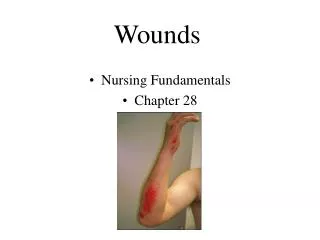
Arterial Ulcers • Characteristics • “punched out” appearance • Smooth wound edges • Surrounding skin may exhibit dusky erythema • Cool to touch • Hairless, thin, brittle with shiny texture
Arterial Ulcers • Typically lower leg • Lateral foot • Toes • Pressure points or where injury has occurred
Arterial Ulcers • Jesse, why do I care what these look like and are you done showing these nasty pictures?
Role of Sonographer • Patient history • Physical exam • ABI’s • Clues to what is going on before you put the transducer on the patient
Role of Duplex • Presence or absence of disease • Severity • Physiologic • Anatomic • Location • Single level • Asymptomatic • claudication • Multi-level • Claudication • Rest pain • ulcerations
Pressures • Ankle/Brachial Index (ABI) • 1.0-1.2 Normal • 0.92-0.99 may indicate presence of arterial obstruction • <0.92 Evidence of arterial obstruction, claudication • <0.40 associated with rest pain or tissue loss
Pressures--Toe • Photoplethysmography (PPG) • Infrared light which responds to changes in blood content near the surface of the skin • Waveform analysis and pressure measurement
Pressures--Toe • Disease from the level of the ankle to the toe • Diabetics • Wound healing potential • Absolute number and index
Pressures--Toe • A toe/ankle index >0.60 suggests the absence of hemodynamically significant obstruction between the ankle and the toe • A toe/brachial index >0.60 suggests the absence of hemodynamically significant obstruction between the heart and the digit
TCPo2 • Transcutaneous oxygen tension • Evaluates oxygen delivery to tissue • Indirect measure of local blood flow • Aids in determining wound healing potential • Patient in supine position • Small electrodes placed at chest, below knee, and 2 over dorsum of foot • Electrodes in the sensors heat area underneath the skin to dilate capillaries • Results recorded and measured in mmHg • >30 mmHg – greater success for wound healing • <30 mmHg - suggests high likelihood of wound not healing
Pressures--Segmental • Typically 3 or 4 cuff system • High thigh, above knee, calf, ankle • Measures pressure at each level • >30mmHg gradient from level to level is significant • >40mmHg indicates occlusion • >20mmHg from side to side is also significant
Pressures--Segmental • Pitfalls include • Medial arterial calcification • Limb girth • Inappropriate cuff size • Can be uncomfortable for patient
Pulse Volume Recordings (PVR) • Measures pressure changes in the bladder of the cuff wrapped around the leg • These changes reflect change in cuff volume • Can use same cuffs as used for segmental pressures
PVR • A 1mmHg pressure change detected in the cuff produces a 20mm deflection (amplitude) on the chart recorder • Using appropriate size cuffs, a preset pressure is obtained • A recording is then obtained
PVR/Segmental Pressures • PVR waveforms and segmental pressures are complimentary tests • If differences exist then a source of error should be investigated
Duplex • Image based evaluation • Looking for anatomic disease and physiologic disease
PW Doppler--Duplex Velocity Ratio = v2/v1 V2= highest peak systolic velocity V1= proximal normal vessel Velocity Ratio (VR) = 6.1
Arterial Ulcers • Role of Duplex essential to understanding presence, location, and severity of disease • Guides intervention and management • Indicator wound healing potential
Venous Ulcers • Result of sustained venous hypertension(Chronic venous insufficiency) • Incompetent valves or poor calf muscle pump • Local venous dilatation and pooling • Traps leukocytes that may release proteolytic enzymes that destroy tissues • May also “trap” important growth factors within vein rendering them unavailable for wound repair
Venous • 70%-90% of chronic wound cases • Estimated 2.5 million patients in the US • Rarely fatal - can severely diminish quality of life
Venous Ulcers • CVI Risk factors • > Age • Hx DVT • Surgery • Restricted mobility • CHF • Cancer • Obesity • Smoking • Family hx VTE • Hypercoable state (Factor V Leiden, Protein C/S deficiency, etc.) • Sedentary lifestyle • Varicosities
Venous Ulcers • Wound characteristics • Gaiter region – medial malleolus • Superficial, irregular shape • Skin shiny and tight (edema) • Brown or purple discoloration – “stasis skin changes”
Varicose Veins • Complications • Swelling • Pain/aching • itching • Leg heaviness • Phlebitis – inflammation of vein • Superficial thrombophlebitis • bleeding • Cosmetic • Not commonly associated with venous ulcers when isolated to the superficial system
Role of the Sonographer • Patient history • Physical exam • Clues to what is going on before you put the transducer on the patient
Role of Duplex • Presence or absence of disease • Severity • Physiologic • Anatomic • Location • Deep • Superficial
Venous Obstruction • Presence or absence of deep or superficial venous obstruction • Compression ultrasound