Download
1 / 34
340 likes | 357 Vues
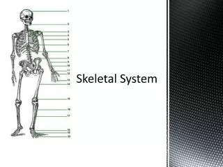
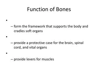
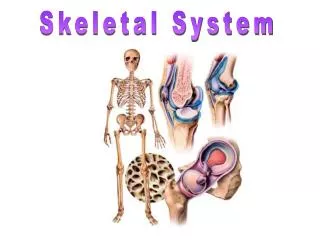
Skeletal System. Function is to: P rovide support & framework for the body P rotec t internal organs Makes movement possible S torage site for minerals Produces blood cells. Skeletal System Intro Video. Bones. Are comprised mostly of phosphorous and calcium & vitamin D

E N D
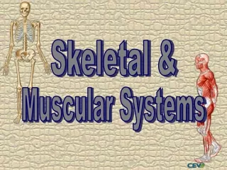
Skeletal System • Function is to: • Provide support & framework for the body • Protect internal organs • Makes movement possible • Storage site for minerals • Produces blood cells Skeletal System Intro Video
Bones • Are comprised mostly of phosphorous and calcium & vitamin D • Are surrounded by the periosteum • thick layer of connective tissue that covers bone • Oxygen and nutrients are carried by blood vessels through the periosteum to the bone osteo- means ?
Bones • Bone marrow is contained within bone cavities • yellow bone marrow • comprised mainly of fat cells • red bone marrow • functions to produce red blood cells, some white blood cells and platelets
Axial skeleton Skull: cranium/ face Vertebral column Thorax: ribs/ sternum Appendicular skeleton Upper Extremities Shoulder girdle, arms, hands Lower Extremities Hip girdle, legs, feet Divisions
How am I able to move my arms and legs? • Bones are attached together by joints • acts similar to a door hinge • some allow movement, while others do not • movement is dependent upon type of joint • immovable • slightly moveable • freely movable
Immoveable Joints • Called fixed joints-- SYNARTHROSIS • Allow for NO movement • Are held together by connective tissue, or are fused together Example: bones in the skull are immovable
Slightly Moveable Joints • Provide for restricted movement-- AMPHIARTHROSIS • Bones are separated from each other Example: joint between two bones in the lower leg (tibia and fibula)
Freely Moveable Joints • Allow for movement in one or more directions-- DIARTHROSIS • Are grouped by type of movement allowed • ball-and-socket- allow for circular movement • hinge- permit back and forth movement • pivot- allow one bone to rotate around another • saddle- permits a bone to slide in two directions
Can you think of an example for each type of joint? • Ball-and-socket • Hinge • Pivot • Saddle shoulder knee Ulna & radius at elbow joint between carpals and metacarpals
Freely Moveable Joints Saddle joint
Are bands of connective tissue connecting bones to bones Are covered with the synovial membrane Are bands of connective tissue connecting muscles to bones Tendon sheaths wrap around tendons that incur a great deal of friction Ligaments Tendons
Types of Fractures • Greenstick: bone is bent and splits; incomplete • Depressed: broken piece moves inward; skull • Compound: ruptures through the skin • Spiral: twisting; results in 1 or more breaks • Impacted: bones jam together • Simple/ closed: complete break with no damage to skin • Comminuted: fragments; breaks in more than 1 piece Reduction: Putting a body part back into proper alignment
greenstick depressed comminuted compound impacted closed spiral
Partner Up! • You can do or say anything to get your partner to say the correct word on the screen. • Keep track of how many you get
Comminuted Impacted Appendicular Cervical Joint Myel(o) Tendon Calcium Osteo Support
Greenstick Ball & Socket Depressed Reduction Protection Hinge Phosphorus Axial Bone Marrow Spiral Fracture
Physicians and Health Care Professionals Orthopedist- medical doctor who treat disorders of the musculoskeletal system Osteopath- physician who combines traditional treatment with manipulative procedures. (hands) Rheumatologist- physician who treat disorders of the joints Podiatrist- medical specialist who treat disorders of the foot Chiropractor- manipulate the spine to treat certain ailments
Laboratory Tests Rheumatoid factor- blood test to confirm rheumatoid arthritis Serum Creatine Phosphokinase (CPK)- high levels appear in some disorders: Brain, muscle tissue or heart damage Serum calcium and serum phosphorus- blood test to determine the body’s calcium and phosphorous levels:Liver disease, cardiovascular disease, osteoporosis Uric acid- high levels lead to gout
Tests and Diagnostic Procedures X-rays- show if bones are broken or not CT Scan- show bone, joint or connective tissue disease Magnetic Resonance Imaging (MRI)- used to detect musculoskeletal system, especially of the soft tissue. Bone Scan -used to detect various bone issues, i.e. tumors • Areas that absorb little or no amount of tracer appear as dark or "cold" spots. This could show a lack of blood supply to the bone or certain types of cancer. • Areas of fast bone growth or repair absorb more tracer and show up as bright or "hot" spots in the pictures. Hot spots may point to problems such as arthritis, a tumor, a fracture, or an infection
Diagnostic Procedures cont. Diskography -exam of the disks by injection, a contrast medium and using radiography. Myelography-radiography of the spinal cord to identify spinal cord conditions. Electromyogram -graphic image of the electrical activity of the muscles. Goniometer -used to measure motion in the joints Densitometer-uses light and x-ray images to measure the density of the bones.
Arthrocentesissurgical puncture to remove fluid from the joint
Arthroscopy Examination with an instrument that explores the interior of a joint.
Diseases of the Skeletal System • Scoliosis: side to side curvature of the back • Sprain: tearing of a ligament; twisting • Bursitis: inflammation of fluid- filled sacs around joints • Osteomyelitis: inflammation of the bone or bone marrow; pathogenic organism • Arthritis: inflammation of joints • Osteoporosis: weakening of bone, caused by hormone deficiency or lack of calcium over a long period of time • Herniated Disc: intervertebral disc protrusion • Dislocation: bone displaced from joint • Kyphosis: curving of the spine which leads to hunchback
Bunion / Bunionectomy Bony bump that forms on the joint at the base of your big toe.
Kyphosis Dislocation Herniated Disc Lordosis Arthritis Gout Scoliosis
Partner Up! • You can do or say anything to get your partner to say the correct word on the screen. • Keep track of how many you get
Comminuted Dislocation Osteopathy Herniated Disc Lordosis CT Scan Scoliosis Axial Osteomyelitis Spiral Fracture
Greenstick Bunionectomy Osteoporosis Kyphosis Arthroscopy Arthritis Appendicular X-Ray Ball & Socket Bursitis