Download
1 / 1
10 likes | 272 Vues
Morphologic Feature on US. L/NL ratio on SMM. Differential Diagnosis of Breast Mass on Ultrasonography & Scintimammography based on Computer aided Diagnosis (CAD). Kyung-hoon Hwang, Jong Hyo Kim * , Jun Gu Lee * , Wonsick Choe and Minki Yoon

E N D
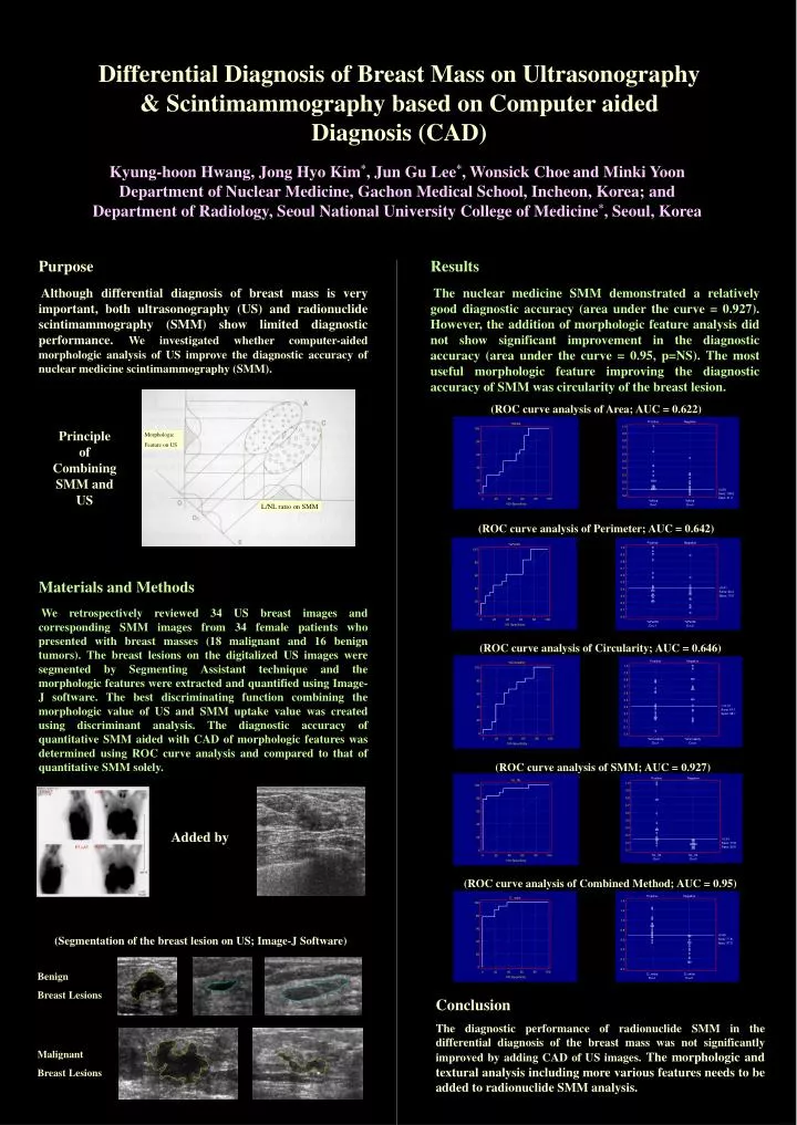
Morphologic Feature on US L/NL ratio on SMM Differential Diagnosis of Breast Mass on Ultrasonography & Scintimammography based on Computer aided Diagnosis (CAD) Kyung-hoon Hwang, Jong Hyo Kim*, Jun Gu Lee*, Wonsick Choeand Minki Yoon Department of Nuclear Medicine, Gachon Medical School, Incheon, Korea; and Department of Radiology, Seoul National University College of Medicine*, Seoul, Korea Purpose Although differential diagnosis of breast mass is very important, both ultrasonography (US) and radionuclide scintimammography (SMM) show limited diagnostic performance. We investigated whether computer-aided morphologic analysis of US improve the diagnostic accuracy of nuclear medicine scintimammography (SMM). Results The nuclear medicine SMM demonstrated a relatively good diagnostic accuracy (area under the curve = 0.927). However, the addition of morphologic feature analysis did not show significant improvement in the diagnostic accuracy (area under the curve = 0.95, p=NS). The most useful morphologic feature improving the diagnostic accuracy of SMM was circularity of the breast lesion. (ROC curve analysis of Area; AUC = 0.622) Principle of Combining SMM and US (ROC curve analysis of Perimeter; AUC = 0.642) Materials and Methods We retrospectively reviewed 34 US breast images and corresponding SMM images from 34 female patients who presented with breast masses (18 malignant and 16 benign tumors). The breast lesions on the digitalized US images were segmented by Segmenting Assistant technique and the morphologic features were extracted and quantified using Image-J software. The best discriminating function combining the morphologic value of US and SMM uptake value was created using discriminant analysis. The diagnostic accuracy of quantitative SMM aided with CAD of morphologic features was determined using ROC curve analysis and compared to that of quantitative SMM solely. (ROC curve analysis of Circularity; AUC = 0.646) (ROC curve analysis of SMM; AUC = 0.927) Added by (ROC curve analysis of Combined Method; AUC = 0.95) (Segmentation of the breast lesion on US; Image-J Software) Benign Breast Lesions Conclusion The diagnostic performance of radionuclide SMM in the differential diagnosis of the breast mass was not significantly improved by adding CAD of US images. The morphologic and textural analysis including more various features needs to be added to radionuclide SMM analysis. Malignant Breast Lesions