Mitotic Cell Division - Exercise 7
Mitotic Cell Division - Exercise 7. Objectives -Know the stages of the cell cycle. -Know why mitosis is important. -Know what types of cells do mitotic division. -Distinguish differences between an animal and a plant cell during mitotic division.

Mitotic Cell Division - Exercise 7
E N D
Presentation Transcript
Mitotic Cell Division - Exercise 7 Objectives -Know the stages of the cell cycle. -Know why mitosis is important. -Know what types of cells do mitotic division. -Distinguish differences between an animal and a plant cell during mitotic division. -Identify the different stages of mitosis under the microscope and in models. NOTE: YOU DO NOT NEED TO IDENTIFY CELLS IN A POLAR VIEW.
Where one chromosomes is double stranded helices replicates so that we now have 2 helices joined by region called center mere or kinetochore and each of these are now called twin chromatidor sister chromatidbecause one DNA double helix replicated itself.
0.5 µm Chromosomes DNA molecules Chromo- some arm Chromosome duplication (including DNA synthesis) Centromere Sister chromatids Separation of sister chromatids Centromere Sister chromatids
MITOSIS is a growth mechanism providing for the production of clone daughter somatic cells and is partially responsible for the processes of embryonic development, growth, and healing. Objective to create two nuclei (cells). Mitosis is to duplicate a set of chromosomes in a parent cell to two daughter nuclei. All cells have identical genome because raise from one fertilize ovule in a process called mitosis. -Duplication of cells (46 chromosomes)-Produces somatic cells
AUTOSOMES • Autosomesthese are the non sex determining chromosomes. These are your cells that are involved in mitotic division, such as, liver, skin, bone cells. • Humans- have a total of 46 chromosomes. Twenty two pairs of autosomes and one pair of sex chromosomes. • Cell division in eukaryotes typically involves the distribution of duplication sets of chromosomes in a parent cell to two daughter nuclei (Mitosis or karyokinesis) and the division of the cytoplasmic contents of a parent cell into two daughter cells (cytokinesis). Cell division is only one stage (the last (4th) stage – M) of the cell cycle. • Mitosis is when nuclear material divides (Division of nucleus) and cytokinesis is when cytoplasm divides.
Interphase • G1: First growth event. The newly formed cell actively produces RNA and protein as it grows – cell growth. • G0: Period of no growth and it occurs prior to S phase, if it is going to happen. • S-phase: Cell stops producing RNA and switches to DNA replication. Chromosomes are duplicated during the S-phase of the cell cycle. Think of S as Synthesis of DNA. • G2: Once the DNA has been duplicated, the cell enters another round of RNA and protein synthesis and growth – more cell growth. • When cells are in G1, S, and G2, they are often said to be in “interphase” – which literally means, between mitotic division. • Finally the cell will divide, and this stage is called M phase. Chromosomes are too thin to see in the interphase stage under the microscope.
THE CELL CYCLE • G1 phase- growth phase where cell makes components for DNA synthesis. • S phase-DNA synthesis, chromosomes replicate. DNA replicates (synthesis) • G2 phase-growth phase where cell makes components for DNA synthesis. • G0 phase-non-dividing cells. Neurons stay in this state. • INTERPHASE-time between cell divisions-everything that is not mitosis. • Mitosis (M-phase)- cell divides: • Prophase • Metaphase • Anaphase • Telophase & Cytokinesis Interphase is usually about 90%, and mitosis is the remaining 10% of the cell cycle.
MITOSIS • A single (2n,diploid) cell divides to form two genetically identical (2n,diploid) daughter cells. • 4 stages: Prophase, Metaphase, Anaphase, &Telophase (Cytokinesis)
Plant cell division • It is necessary to obtain tissues that contain cells that are actively dividing. One such tissue is the root tip. • Note: The viewpoint your looking at cells is called a lateral view. • Each cell can be considered to be a single frame of a motion picture. Also know the parts of the cells when dividing. • Onion root tip (Alliumcepa). Most of the cells you see will not be in the M stage of the cell cycle. That is, most cells will be in interphase. Animal Cell Division Mitosis is animal cells in most readily seen in early embryonic stages. The blastula stage of an embryo is more or less a spherical ball of cells, and cell division occurs in every imaginable plane of the embryo. Whitefish blastula.
NOTE!!! • A professor could ask you what are the different stages of mitosis? In that case, you would write down prophase, metaphase, anaphase, and telophase. • A professor could ask you the different stages of the cell cycle? In that case, you would include interphase (G1, S, G2), and mitosis (P,M,A,T).
Question How can you tell whether you’re looking at a plant cell or an animal cell?
Growth period: production of proteins and cytoplasmic organelles
INTERPHASE • The only significant genetic event occurring here is the replication of DNA (all the chromosomes duplicate). • In interphase the chromosomes are not visible. They exist in thread-like strands known as CHROMATIN • Cell is not diving. Very prominent nucleus (no chromosomes) cell makes protein and lots of RNA.
Interphase These cells have obvious nuclear membranes and the nuclear material is still dispersed as chromatin.
PROPHASE CELLS When mitosis begins, several changes occur in the cell. The chromosomal material (called chromatin) in the nucleus starts to condense, and eventually reaches thickness that makes them visible under the light microscope. As prophase proceeds, the chromosomes continue to get shorter and thicker, and eventually begin to move toward the equator of the cell, midway between the poles. Prophase is typically the longest phase of mitosis. interphase prophase -Chromosomes condense and become visible -Nuclear membrane breaks down -Nucleoli vanish
Prophase Several cells are seen in various stages of prophase. Note the presence/absence of a nuclear membrane and the spindle apparatus, and the chromatin beginning to condense as visible chromosomes.
METAPHASE CELL Chromosomes are short and thick will reach the center of the cell called the equatorial plane or metaphase plate. The duplicated chromosomes are separated from one another, except at one point called the kinetochore (centromere). When the duplicate chromosomes are in this attached condition, as they have been since they were duplicated, each strand is called a sister chromatid. At the conclusion of metaphase each centromere, holding two identical sister chromatids, is attached to two spindle fibers, one from each pole. -chromsomes line up on the center line of the cell -centromeres duplicate -spindle fibers attach to centromeres
Metaphase The two cells in the center are in metaphase. In each, notice the chromosomes are aligned at the cell’s equator and the well formed spindle apparatus.
ANAPHASE CELL Anaphase begins with the separation of sister chromatids of each duplicated chromosome. Each chromatid is moved by motor molecules that ratchet along the spindle fibers toward the polar ends. The original chromosome and its exact duplicate are pulled to opposite poles of the cell. This happens for each different chromosome that the cell possesses; thus, each pole will eventually obtain exactly the same set of chromosomes that were in the original nucleus. In anaphase the chromatids are separated. • -Sister chromatids move to opposite poles of the cell • -Spindle fibers pull the chromosomes apart by the centromeres
Early Anaphase The spindle apparatus has begun to separate the chromosomes in the upper cell. The cell near the bottom of the field as in metaphase.
Late Anaphase • The chromosomes are close to the centrosomes, which means they have moved apart just about as far as they will go.
TELOPHASE CELL Plant cell Animal cell The events of telophase are essentially the reverse of the those of prophase. At the end of this, there are two nuclei, each having identical sets of chromosomes.
TELOPHASE • Sister chromatids reach the opposite poles and begin returning to the interphase state. • Nuclear membrane and nucleoli reform. • Spindle fibers disperse. • Cytokinesis occurs. • A cell plate (plants) or a cleavage furrow (animals) forms.
Cytokinesis : division of cytoplasm • Animal cells : cleavage • Cleavage furrow • Division begin as a shallow groove in the cell surface near the metaphase plate • Contractile ring (on the cytoplasmic side of the furrow) • Composes of actin microfilaments and the protein myosin • Cleavage furrow deepens until the parent cell is pinched in two • Plant cells : cell plate formation • Vesicles from the Golgi collect at the middle of the cell producing a cell plate • Cell wall material carried in the vesicles is deposited on the plate as it grows, until it fuses with the membrane along the perimeter of the cell
Late Telophase and Cytokinesis The cleavage furrow has nearly divided the two cells, so cytokinesis is almost complete. Telophase will continue with the chromosomes dispersing as chromatin and the nuclear envelope reforming.
Polar view The cell in the center doesn’t fit any of the description because it is begin viewed from one end (pole) rather than from above.
Cleavage furrow The cleavage furrow (CF) has begun to form.
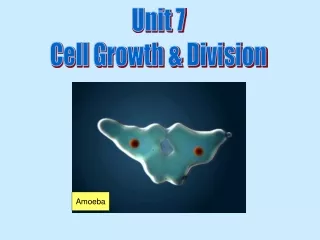
Cell Division A single-celled eukaryote (amoeba) reproduces • Reproduction • Equal distribution of genetic material to two daughter cells • Growth • Sexually reproducing organisms develop from a single cell (zygote) • Repair • Replace cells that die from normal wear and tear or accidents Sand dollar embryo after the egg divided to form 2 cells Dividing bone marrow cells produce new blood cells
Animal Vs. Plant Cells -What are some differences between an animal cell and a plant cell?
Animal Vs. Plant Cells -What are some differences between an animal cell and a plant cell? Plants have a cell wall, after telophase plant cells divide by forming a cell plate, and animal cells form a cleavage furrow. Plant cells are rectangular, and animal cells are more circular.
Page 8 – Lab Book • 1. Why are the onion root tip and the whitefish blastula useful tissues for the study of cell division? • 2. Distinguish between mitosis and cytokinesis. • 3. If a cell has 16 chromosomes when it is in G1, how many chromosomes will there be in each daughter cell following a mitotic cell division? • 4. What are the genetic consequences of mitotic cell division for the resulting daughter cells? • 6. How do plant and animal cells differ in the execution of cytokinesis? Why don’t plant cells undergo cytokinesis in the same manner as animal cells?