SPINAL CORD, SPINAL NERVES, EYE AND EAR ANATOMY
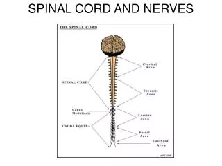
SPINAL CORD, SPINAL NERVES, EYE AND EAR ANATOMY. Learning Objectives. Identify structures of the spinal cord, both gross anatomical structures and cross sectional structures. Identify meninges and spaces around the spinal cord. Identify major peripheral nerves and nerve plexuses.

SPINAL CORD, SPINAL NERVES, EYE AND EAR ANATOMY
E N D
Presentation Transcript
Learning Objectives • Identify structures of the spinal cord, both gross anatomical structures and cross sectional structures. • Identify meninges and spaces around the spinal cord. • Identify major peripheral nerves and nerve plexuses. • Identify the accessory and internal structures of the eye, and explain their function. • Describe the structures of the external and middle ear and explain how they function.
Spinal Nerves: Roots Figure 13.7a
Spinal Nerves Figure 13.6
Nerve Plexuses • All ventral rami except T2-T12 form interlacing nerve networks called plexuses • Plexuses are found in the cervical, brachial, lumbar, and sacral regions • Each resulting branch of a plexus contains fibers from several spinal nerves
Cervical Plexus Figure 13.8
Brachial Plexus Figure 13.9a
Brachial Plexus: Distribution of Nerves Figure 13.9c
Spinal Nerve Innervation: Back, Anterolateral Thorax, and Abdominal Wall Figure 13.7b
Lumbar Plexus Figure 13.10
Sacral Plexus Figure 13.11
The eye • Conjunctiva covers most of eye • Cornea is transparent anterior portion • Three layers • Outer fibrous tunic • Sclera, cornea, limbus • Middle vascular tunic • Iris, ciliary body, choroid • Inner nervous tunic • Retina