GAIT DISTURBANCES
GAIT DISTURBANCES. Anshul Jain. Outline. Hemiplegic gait Diplegic Gait Parkinsonian Gait Myopathic Gait Ataxic Gait Choreiform Gait (Hyperkinetic Gait) Sensory Gait Neuropathic Gait ( Steppage Gait, Equine Gait ). Facts.

GAIT DISTURBANCES
E N D
Presentation Transcript
GAIT DISTURBANCES Anshul Jain
Outline • Hemiplegic gait • Diplegic Gait • Parkinsonian Gait • Myopathic Gait • Ataxic Gait • Choreiform Gait (Hyperkinetic Gait) • Sensory Gait • Neuropathic Gait (Steppage Gait, Equine Gait)
Facts • Gait and balance disorders and weakness is the 2nd most common cause of falls in older persons. • Gait speed declines @ 12-16% per decade after age 60 years • Slow walkers ( < 0.6 m/sec ) • Fast walkers ( > 1 m/sec ) • Gait speed < 0.6 m/sec predicts early mortality
Hemiplegic gait • Upper motor neuron weakness secondary to stroke • The patient stands with unilateral weakness on the affected side, arm flexed, adducted and internally rotated. Leg on same side is in extension with plantar flexion of the foot and toes. • When walking, the patient will hold his arm to one side and drags his or her affected leg in a semicircle (circumduction) due to weakness of distal muscles and extensor hypertonia in lower limb.
Diplegic Gait • Bilateral periventricular lesions ex cerebral palsy • Patients have involvement on both sides with spasticity in lower extremities worse than upper extremities. The patient walks with an abnormally narrow base, dragging both legs and scraping the toes. • There is also characteristic extreme tightness of hip adductors which can cause legs to cross the midline referred to as a scissors gait.
Parkinsonian Gait • This type of gait is seen with rigidity and hypokinesia from basal ganglia disease • The patient's posture is stooped forward with flexion at the knees. • Gait initiation is slow and steps are small and shuffling. Arm swing is reduced. • Turning is en bloc like a statue. • The patient may show an involuntary inclination to take accelerating steps, known as festination.
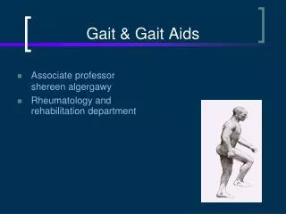
Myopathic Gait • This happens secondary to hip abductor weakness. Can be seen in myopathies like muscular dystrophy • Hip girdle muscles are responsible for keeping the pelvis level when walking. If someone have weakness on one side, this will lead to a drop in the pelvis on the contralateral side of the pelvis while walking (Trendelenburg sign). • With bilateral weakness, you will have dropping of the pelvis on both sides during walking leading to waddling.
Ataxic Gait • Most commonly seen in cerebellar disease, this gait is described as clumsy, staggering movements with a wide-based gait. • While standing still, the patient's body may swagger back and forth and from side to side. Patients will not be able to walk from heel to toe or in a straight line. • The gait of acute alcohol intoxication will resemble the gait of cerebellar disease.
Choreiform Gait (Hyperkinetic Gait) • This gait is seen with certain basal ganglia disorders including Sydenham's chorea, Huntington's Disease and other forms of chorea, athetosis or dystonia. • The patient will display irregular, jerky, involuntary movements in all extremities. • Walking may accentuate their baseline movement disorder.
Sensory Gait • This gait can be seen in disorders of the dorsal columns (B12 deficiency or tabesdorsalis) or in diseases affecting the peripheral nerves (uncontrolled diabetes). • Normally when our feet touch the ground, we receive propioreceptive information to tell us their location. The sensory ataxic gait occurs when there is loss of this propioreceptive input. In an effort to know when the feet land and their location, the patient will slam the foot hard onto the ground in order to sense it. A key to this gait involves its exacerbation when patients cannot see their feet (i.e. in the dark). • This gait is also sometimes referred to as a stomping gait since patients may lift their legs very high to hit the ground hard. In its severe form, this gait can cause an ataxia that resembles the cerebellar ataxic gait.
Neuropathic Gait (Steppage Gait, Equine Gait) • Seen in patients with foot drop (weakness of foot dorsiflexion), the cause of this gait is due to an attempt to lift the leg high enough during walking so that the foot does not drag on the floor. • If unilateral, causes include peroneal nerve palsy and L5 radiculopathy. • If bilateral, causes include amyotrophic lateral sclerosis, Charcot-Marie-Tooth disease and other peripheral neuropathies including those associated with uncontrolled diabetes.