Download
1 / 18
190 likes | 370 Vues
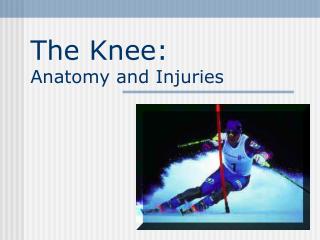
Learn about knee anatomy, medial and lateral condyles, ligaments like MCL and ACL, meniscus, tests for instability and injuries, and joint space orientation. Discover mechanisms of injury and how to detect and treat knee ligament sprains.

E N D
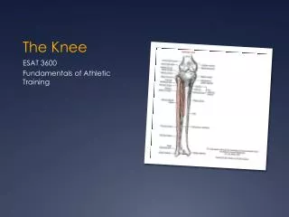
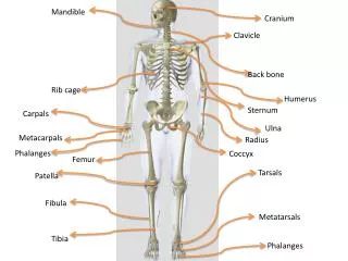
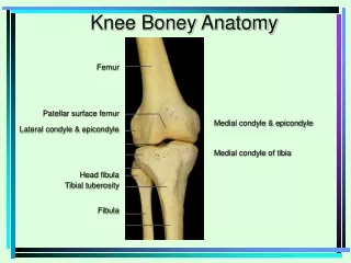
Knee Boney Anatomy Medial condyle & epicondyle Medial condyle of tibia Femur Patellar surface femur Lateral condyle & epicondyle Head fibula Tibial tuberosity Fibula
MCL Mechanism of Injury Valgus Stress MCL Sprain 1st Degree 2nd Degree 3rd Degree
Valgus Stress Test Stresses MCL Valgus Stress at 0 - 5º Valgus Stress at 25 - 30 º
MOI of LCL Injury Varus Stress Test Stresses lateral structures Varus Stress Varus Stress at 0 º and 25 º to 30º of flexion
ACL Tear • Anterior instability • Mechanism • Deceleration injury • IR of femur with knee flexed and foot planted • Hyperextension of knee • Swelling • Pop at time of injury • Pain with • AROM • PROM • Anterior instability • Decreased strength • Giving way or buckling Signs and Symptoms
Anterior Drawer Test Grading Anterior InstabilityMedial viewRight knee • Stabilize Foot • Check for hamstrings relaxation • Thumbs either side patellar tendon • Apply anterior force • Grade amount of translation
Lachman’s Test • Better test than Anterior Drawer • Takes opposition of hamstrings out of play • Knee flexed 15 º - 30º • Stabilize femur • Apply anterior force to tibia
Pivot Shift Test • Gold standard test for ACL • Leg is externally rotated • Valgus force is applied as leg is flexed • Positive test indicated by clunk sensation
Posterior CruciateLigament Posterior cruciate
Posterior Sag Test • Posterior Cruciate vs Anterior Cruciate • Athlete supine • Both knees flexed 90’ • Observe laterally
Posterior Drawer Test • Athlete supine • Knee flexed 90’ • Foot neutral • Sit on foot to stabilize it • Posterior force applied at tibial plateau • Positive test indicates PCL injury PCL ACL
Medial and Lateral Meniscus Medial meniscus “C” shaped Lateral meniscus more circular shaped • Mechanism of Injury • Squat with rotation • Internal rotation of femur • on fixed tibia
Joint Space Orientation Lateral Meniscus Medial Meniscus Medial Joint Space Lateral Joint Space
Mc Murray Test • Flex knee fully • Palpate medial & lateral joint spaces with one hand • Rotate tibia opposite to femur as knee is extended • Palpable pop and/or pain indicate a positive test
Apley’s Compression Test External rotation of tibia tests medial meniscus Internal rotation of tibia tests lateral meniscus Apley’s Distraction Test Unloads the meniscus Stressess MCL and LCL