Download

1 / 45
490 likes | 603 Vues
Learn about the antigens associated with immunologically mediated renal diseases, including mechanisms of Type I-III hypersensitivity, cell-mediated immunity, and complement activation. Discover how immune complexes and cytotoxic antibodies lead to renal damage. Uncover the role of different immune cells in glomerular injury and the inflammatory response in renal diseases.

E N D
Immunologic mechanisms of renal diseases Qingqing Wang, Professor Institute of Immunology, ZJU Email: wqq@zju.edu.cn
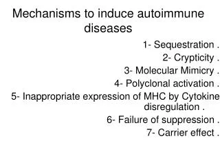
Antigens The cause of immunologically mediated renal disease is antigenic triggering of an immune reaction. The list of associated antigens is extensive and continually expanding. These antigens are categorized as renal or non-renal and as self or foreign . The causative antigen is often unknown.
Antigens associated withimmunologically mediated renal disease
Antigens To cause immunologically mediated renal disease, an antigen must localize to the kidney and trigger a local immune inflammatory response. Renal antigens are inherently localized, being constituent proteins of the kidney. Non-renal antigens require a mechanism for depositing in the kidney.
Immunologic mechanisms of renal diseases Type I hypersensitivity (IgE) Type II hypersensitivity (Cytotoxic Antibody-mediated ) Type III hypersensitivity (Immune Complex-mediated ) Cell-mediated immunity (CD4+, CD8+ T) Complement activation Immune hereditary factors (HLA)
Type II hypersensitivity Ags on the surface of target cells ↓ body→IgG, IgM ↓ 1. damage the target cell 1) activation of complements 2)opsonization: FcR C3bR 3) neutrophil activation 2. target cell dysfunction
Cytotoxic Antibody-mediated Renal Disease Prototype: Anti-GBM disease (Goodpasture's disease) 抗基膜性肾小球肾炎 Renal damage is caused by linear deposition of antibody specific for type IV collagen α3 chain of the GBM. The antibody attaches to its antigen and activates the complement. Cytotoxic antibody localizes along the GBM in a linear pattern with C.
Goodpasture syndrome: 肺出血 & 肾小球肾炎
连续线形荧光 肾小球基底膜抗原 glomerular inflammation and severe damage
Type III hypersensitivity Ag→body→IgG, IgM, IgA ↓ immune complexes (IC) ↓ soluble IC ↓ ICs are deposited from the circulation into vascular basement membranes ①↓ ② ↓FcR activation of complement plat. and basophils ↓ C3a, C5a →mast cell → release of vasoactive amines ↓ basophils ③ Neutrophils vasodilation ↓ lysosomal edema enzymes→damage the tissue
Immune Complex-mediated Renal Disease Planted antigen attracts its antibody from the circulation, and a local immune complex is formed. Immune complex localizes in the mesangium, glomerular capillary wall, or renal interstitium as a lumpy-bumpy pattern. Small immune complexes are less likely to be deposited, and large immune complexes are preferentially removed by RES minimizing localization in the kidney. As circulating immune complexes are formed and antibody production increases, the size of the circulating immune complex increases: • removal from the circulation by RES cells or • localization in the mesangium or glomerular capillary wall. Various endogenous and exogenous substances may function as antigen in immune complex formation. • endogenous nuclear proteins in DNA-anti-DNA IC in lupus nephritis, streptococcal cell wall antigens in post-streptococcal glomerulonephritis.
Epithelial cell injuryThe postulated sequence is a consequence of antibodies against epithelial cell antigens, toxins, cytokines, or other factors causing injury with foot process effacement and sometimes detachment of epithelial cells and protein leakage through defective GBM and filtration slits.
Cells involved glomerular injury 1 Neutrophils and monocytes infiltrate the glomerulus in certain types of glomerulonephritis, largely owing to activation of complement, resulting in generation of chemotactic agents (mainly C5a), but also by Fc-mediated adherence and activation. Neutrophils release proteases, which cause GBM degradation; oxygen-derived free radicals, which cause cell damage; and arachidonic acid metabolites, which contribute to the reductions in glomerular filtration rate (GFR). 2 Macrophages, T lymphocytes, and natural killer (NK) cells, which infiltrate the glomerulus in antibody- and cell- mediated reactions, when activated release a vast number of biologically active molecules.
Cells involved in glomerular injury 3 Platelets aggregate in the glomerulus during immune-mediated injury. Their release of eicosanoids and growth factors may contribute to the manifestations of glomerulonephritis. Antiplatelet agents have beneficial effects in both human and experimental glomerulonephritis. 4 Resident glomerular cells, particularly mesangial cells, can be stimulated to produce several inflammatory mediators, including reactive oxygen species, cytokines, chemokines, growth factors, eicosanoids,nitric oxide, and endothelin. In the absence of leukocytic infiltration, they may initiate inflammatory responses in the glomerulus.
Soluble mediators Complement components induce leukocyte influx and lead to formation of MAC. Pro-inflammatory cytokines, IL-1 and TNF, induce leukocyte adhesion. Chemokines such as MCP-1 and RANTES promote monocyte and lymphocyte influx. Growth factors, PDGF are involved in mesangial cell proliferation.TGF-β and fibroblast growth factor appear to be critical in the ECM deposition and hyalinization leading to glomerulosclerosis in chronic injury. The coagulation system is also a mediator of glomerular damage. Fibrin is frequently present in the glomeruli in glomerulonephritis, and fibrin may leak into Bowman space, serving as a stimulus for parietal epithelial cell proliferation (crescent formation).
Mechanisms of chronic tubulointerstitial injury in glomerulonephritis. Various components of the protein-rich filtrate and cytokines derived from leukocytes cause tubular cell activation and secretion of cytokines, growth factors, and other mediators. These, together with products of macrophages, incite interstitial inflammation and fibrosis. ET-1, endothelin-1, PAI-1, plasminogen activator inhibitor-1; TIMP-1, tissue inhibitor of metalloproteinases.
Immune complex glomerular disease Most patients with lupus nephritis have an immune complex-mediated glomerular disease The standard classification divides these disorders into five different patterns in which (type I) represents no disease • Mesangial (type II) • Focal proliferative (type III) 局灶性 • Diffuse proliferative (type IV) 弥散 • Membranous (type V) 终末期
Renal manifestations of SLE Renal involvement is common in SLE An abnormal urinalysis is present in approximately 50% of patients at the time of diagnosis and eventually develops in more than 75 percent of cases The most frequently observed abnormality is • proteinuria (80 %) • 40% have hematuria and/or pyuria sometime during the course of their illness 血尿,脓尿
‘Bumpy’ appearance of immune complexes deposited in the glomerulus in SLE
ACUTE NEPHRITIC SYNDROME(Acute Glomerulonephritis; Postinfectious Glomerulonephritis) The prototype of an acute nephritic syndrome is poststreptococcal glomerulonephritis (PSGN) due to infection with certain nephritogenic strains of group A -hemolytic streptococci, such as type 12 (associated with pharyngitis咽炎) and type 49 (associated with impetigo脓疱病). Immunofluorescence microscopy usually shows immune complex deposition with IgG and C in a granular pattern. The presenting manifestations range from asymptomatic hematuria (in about 50%) and mild proteinuria to full-blown nephritis with gross or microscopic hematuria proteinuria, oliguria, edema, hypertension, and renal insufficiency.
IgA NEPHROPATHY • Berger's disease is now used to describe any idiopathic IgA nephropathy • Patients have gross or microscopic hematuria, often with high blood pressure. The disease usually runs a chronic, slowly progressive course • Mesangial and focal-segmental proliferation and sclerosis may be seen by light microscopy. In bad cases, crescents develop. • Immunofluorescence shows IgA deposited in the mesangium (often with IgG, IgM, and/or C3, but no C4, i.e., the alternate pathway of complement is being activated.) • Serum IgA is often elevated, and IgA-containing immune complexes are often demonstrable, whether or not there is some primary disease to explain their presence
Cell-mediated Renal Disease The prototype is the renal transplant. In nearly all non twin transplants, the kidney presents nonself antigens that trigger an immune (predominantly cell-mediated) response. If the host has been presensitized to antigens of the renal graft, transplantation may trigger hyperacute rejection , resulting in acute renal ischemia, infarction, and transplant loss. Cell-mediated renal disease appears to play a part in chronic poststreptococcal glomerulonephritis (PSGN). Lymphocytes stimulated by exposure to streptococcal wall antigens may cross-react with renal glomerular antigens, resulting in progressive cell death and sclerosis of the renal parenchyma.
Complement activation Alternative pathway :C3 MPGN(membranoproliferative glomerulonephritis) 膜增生性肾小球肾炎 Ⅰ--- C3 deposit、antibody Ⅱ--- dense deposit Ⅲ--- both above
Immune hereditary factors PSGN has been associated with HLA-B12 IgA nephropathy with HLA-B35 and HLA-DR4 Anti-GBM or Goodpasture's syndrome with HLA-DR2 IMN(idiopathic membeanous nephropathy)with HLA- II(DR3、DR2、DQ2、DQ1) Minimal change nephrosis with HLA-DR7、DR9
Diagnosis Renal biopsy and light microscopic examination of stained tissue provide the best method for diagnosing immunologically mediated renal disease, assessing prognosis, and selecting treatment. Immunofluorescence microscopy using fluorescein-labeled specific antibodies often is also helpful in characterizing the type and location of immune components in the kidney. The type and pattern of C deposition help diagnosis. C deposition usually follows the pattern of immune complex or immunoglobulin deposition or both. However, C3 deposition in the absence of immunoglobulin, Clq, or C4 deposition may occur via alternative pathway activation in type II MPGN.
Serologic Analyses Detect • cytotoxic antibodies in type II renal disease (eg, anti-GBM antibodies, anti-HLA antibodies). • CIC may be found in various immune complex-mediated renal diseases. • Circulating ANCA can be detected in ANCA-mediated renal disease . Altered levels of C proteins often differentiate types of immunologically mediated renal disease. • When alternative pathway activation predominates (eg, in MPGN and frequently PSGN), C consumption begins with activation of C3; thus, early components of C (Clq, C4, and C2) are not depressed. • In classic pathway activation (eg, in SLE), consumption begins with the early components, which are thereby depressed. • The presence of C3 nephritic factor with depressed C3 but normal Clq, C4, and C2 is virtually diagnostic of MPGN with alternative pathway activation.
Histocompatibility testing May help diagnose forms of immunologically mediated renal disease. 链球菌感染后肾小球肾炎 • PSGN has been associated with HLA-B12, • IgA nephropathy with HLA-B35 and HLA-DR4, and • Anti-GBM or Goodpasture's syndrome with HLA-DR2.
Thanks! Contact: 王青青13606640030, wqq@zju.edu.cn