Download
1 / 24
291 likes | 819 Vues
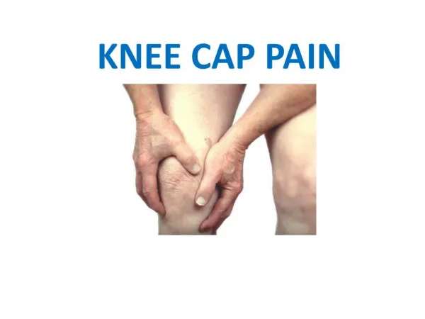
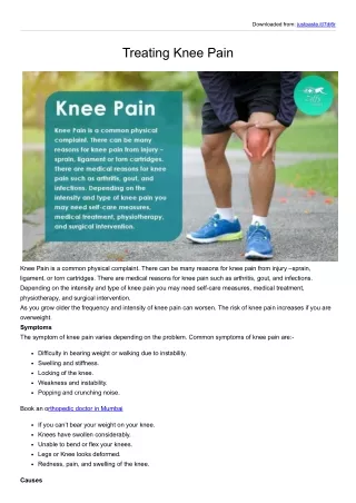
Lateral, Medial and Posterior Knee Pain. Lateral, Medial and Posterior Knee Pain. Knee pain is one of the most common musculoskeletal complaints. Knee joint is tough and once injured unless it is treated correctly, can be difficult to heal.

E N D
Lateral, Medial and Posterior Knee Pain Knee pain is one of the most common musculoskeletal complaints. Knee joint is tough and once injured unless it is treated correctly, can be difficult to heal. Knee pain can be related tooveruse, where small stresses are placed in a large number of times on knee without allowing adequate recovery. For example running too much too soon, or excessive jumping. Or knee pain can be acute where the injury is caused by an impact or twisting of the knee.
Lateral Knee Pain • Iliotibial band friction syndrome (ITBFS); • Excessive lateral pressure syndrome; • Biceps Femoris Tendinitis; • Superior tibiofibular joint injury; • Patellofemoral Pain Syndrome; • Osteoarthritis;
Iliotibial band friction syndrome (ITBFS): Mechanism: Repeated knee flexion/extension, the iliotibial band (ITB) rubs against the prominent lateral epicondyle of the femur. • Cause: The two main causes of ITBFS are- • inappropriate training and abnormal biomechanics. • Excessive downhill running or running on an uneven surface.
Sign & Symptoms: • Pain at or around the lateral epicondyle of the femur. • Pain normally aggrevated by running, • Pain during flexion or extension of the knee. • Decreased strength in hip abduction. • Tenderness present in the gluteal area. • Tightness of the iliotibial band. Treatment: • Rest- avoid painful stimuli, • If swelling is present,the R.I.C.E. technique maybe used. • ITB stretches can be very effective in preventing reoccurring pain. • Self-massage techniques can also be very helpful in correcting excessive ITB tightness. • Perform soft tissue or deep friction massage. • Electrotherapeutic treatment techniques such as TENS, ultrasound and/or interferential Therapy.
Specialist Stretching Techniques Sports Massage Stretches of the right iliotibial band
Excessive lateral pressure syndrome Excessive lateral-pressure syndrome (lateral patellar compression syndrome) occurs with excessive pressure on the lateral patellofemoral joint resulting from a tightlateral retinaculum. This excessive pressure may lead to increased bone strain on the lateral patella, inflammation of the lateral retinaculum and ITBFS. Treatment: • patellofemoral mobilization and soft tissue therapy to the lateral retinaculum. • Taping techniques rarely help. • In more advanced stage, surgical lateral retinacular release, or removal of the lateral patellar fragment, may be required.
Biceps Femoris Tendinitis • Biceps femoris tendinitis occurs with excessive acceleration and deceleration activities. • Sign & Symptoms: • The pain can be produced with resisted flexion, especially with eccentric contractions. • It is often found in association with tightness of the hamstring muscles. Treatment: • Treatment is based on the general principles of the treatment of tendinitis: • reduction of inflammation, • soft tissue therapy, • stretching and strengthening,especially eccentric strengthening.
Soft tissue therapy Eccentric strengthening exercise
Superior tibiofibular joint injury Superior tibiofibular joint injury may result from direct trauma or in association with rotational knee or ankle injuries. Sign & Symptom: Pain is produced by activities demanding tibial rotation. The patient may feel the pain distally in the shin and not localized. On examination, the joint is tender and there may be either restricted or excessive movement on passive gliding of the superior tibiofibular joint. Treatment: • Mobilization. • Local electrotherapy can be used. • Strengthening of tibial rotators. • patients who fail to respond to conservative management, a corticosteroid injection may be used.
Medial Knee Pain Pain about the medial knee is less common than Posterior and lateral knee pain. • Causes: • Patellofemoral syndrome (common); • Synovial plica; • Pes anserinus Tendinitis\Bursitis; • Pellegrini-Stieda syndrome; • Medial meniscus Minor tear\Degenerative change;
Patellofemoral Syndrome In most cases of medial knee pain, the pain is actually anteromedial. most frequently due to patellofemoral syndrome. Sign & Symptom: • Aching pain occurs in the knee joint, particularly at the front, around and under the patella. • Pain under the patella when bending and straightening the knee • .Tenderness along the inside border of the kneecap. • Usually swelling is present. • often worse when walking up or down hills or stairs. • A clicking or cracking sound may be present on bending the knee. • Sitting for long periods may be uncomfortable. • Wasting (atrophy) of the quadriceps muscles. • Tight muscles including calf muscles, hamstrings, quadriceps and iliotibial band.
Treatment: • RICE (Rest, ice compression and elevation) to the injured knee. This will help reduce swelling. • Rest until there is no pain. • Use a knee support or heat retainer (with a hole). • Electrotherapy such as ultrasound, laser and electrical stimulation. • Comprehensive rehabilitation program in conjunction with taping techniques. • Vastus Medialis Obliquus (VMO) strengthening exercises combined with iliotibial band (ITB) stretches. Taping for patella pain Strengthening exercises
Pes anserinus Tendinitis\Bursitis The pes anserinus ('goose's foot'): It is the combined tendinous insertion of the sartorius, gracilis andsemitendinosus tendons at their attachment to the tibia. A bursa, the pes anserinus bursa, lies between this insertion and the bone. The pes anserinus tendon attachment and its associated bursa may become inflamed as a result of overuse in swimmers,cyclists or runners. • Sign\Symptoms • Characterized by localized tenderness and swelling. • Pain may be elicited on active contraction or stretching of the involved muscles. Treatment • Follows the general principles of tendinitis/ bursitis management. • Cortico-steroid injection into the bursa may be extremely effective.
Pellegrini-Stieda syndrome It is a disruption of the femoral origin of the MCL with calcification at the site of injury. It may occur following direct trauma or, less frequently, following grade II or III sprain of the MCL. Sign\Symptoms: It is an important cause of knee stiffness. The patient complains of difficulty straightening the leg and twisting. On examination, there may be a marked restriction in joint range of motion. Treatment: • Active mobilization of the knee joint • Infiltration of a corticosteroid agent to theMCL attachment if inflammation persists.
Posterior knee pain Posterior knee pain is a common site of referred pain from the lumbar spine and from the patellofemoral joint. Alternatively, local structures (e.g. popliteus, hamstring tendon) may cause posterior knee pain. • Causes: • Knee joint effusion • Referred pain from Lumbar area; • Popliteus tendinitis; • Gastrocnemius tendinitis; • Hamstring tendinitis; • Baker's cyst; • Deep venous Thrombosis;
Popliteus tendinitis Pain may arise from the popliteus muscle, its tendon or the popliteus/arcuate ligament. These structures are situated close together and pain in this area is difficult to isolate. Occasionally, it is inflammed as a direct result of overuse in acceleration/deceleration activities. Treatment: • Reduction of the inflammatory process using NSAIDs, • electrical stimulation and ultrasound • Soft tissue therapy andmobilization may help to correct any restriction of tibial rotation, knee flexion. • Posterior knee structures, especially the hamstring muscles, should be stretched. • Strengthening of tibial rotators and hamstring muscles is also necessary.
Strengthening of tibial rotators. This may be performed against manual resistance
Gastrocnemius tendinitis Inflammation of the origin of the medial gastrocnemius at the posterior femoral condyle occurs occasionally with overuse. It may occur as a result of excessive hill running. Sign\Symptoms: • On examination, local tenderness may be elicited. • Pain may be reproduced on resisted knee flexion, jumping, hopping or, occasionally, with stretch of the gastrocnemius muscle. Treatment: • ice, • electrotherapy • NSAIDs to settle inflammation, • soft tissue therapy to correct generalized or focal abnormalities of the gastrocnemius muscle, • graduated stretching/strengthening program.
Hamstring tendinitis The hamstring muscles consist of the Biceps femoris, Semitendinosus and Semimembranosus. These muscles are used to bend (flex) the knee. Inflammation of these muscles can result from a partial rupture that has not healed properly or through overuse, (particularly acceleration and deceleration) Sign & Symptom: • Tenderness and swelling where the tendon inserts onto the bone (tendonitis). • Pain when try to bend the knee against resistance. • Stiffness after exercise. Hamstring tendon inflammation
Treatment: • Rest and apply ice / cold therapy. • anti-inflammatory medication such as ibuprofen. • ultrasound or laser treatment. • Prescribe a full rehabilitation programme consisting of pain reduction, stretching, strengthening and sports massage techniques. Sports Massage
Baker's cyst Baker's cyst is a chronic effusion that herniates between the two heads of the gastrocnemius. It usually occurs secondary to damage to the knee joint, most commonly degeneration or meniscal damage. • Sign & Symptom: • A rounded swelling,the size like a golf ball. • A sensation of pressure in the back of the joint which can go down into the calf muscle. • Difficulties in bending the joint. • Aching and tenderness after exercise. Baker's Cyst / Popliteal Cyst
Treatment: • Rest. • Operate to correct or remove the bursa. Patient should be out of action for 8 to 12 weeks following surgery.
Deep venous thrombosis Deep Vein Thrombosis is a blood clot in a vein. It is more common in the calf muscle area, particularly following surgey. • Sign & Symptom: • Constant calf pain. • Tenderness at a point deep in the muscle. • Swelling. • Increased temperature. • If the ankle is dorsi flexed (toes pushed upwards to stretch the muscle by the therapist whilst the athlete remains relaxed) this may cause pain.