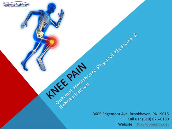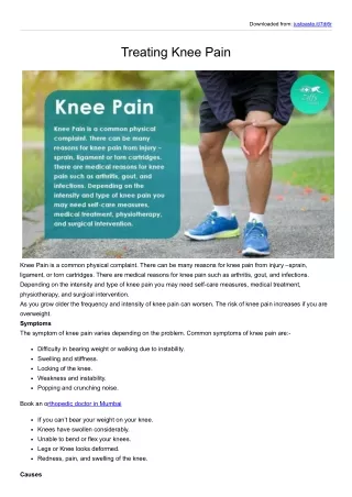
Knee Pain
Knee Pain . Knee Problems for Primary Care. Authors: Margaret Crook, M.D. Editor: Amy W. Shaheen, M.D. Knee Pain. Present in up to 20% of adult population 6% of patients presenting to adult primary care with a physical complaint have knee pain

Knee Pain
E N D
Presentation Transcript
Knee Problems for Primary Care • Authors: Margaret Crook, M.D. • Editor: Amy W. Shaheen, M.D.
Knee Pain • Present in up to 20% of adult population • 6% of patients presenting to adult primary care with a physical complaint have knee pain • The description of knee pain can help narrow the differential diagnosis www.knee-pain-management.com/knee_anatomy.html
Most common diagnoses of knee pain in primary care • Sprains & strains (prevalence 42%) • Osteoarthritis (34%) • Meniscal injuries (9%) • Collateral ligamentous injuries (7%) • Cruciate ligamentous injuries (4%) • Gout (2%) • Fracture (1.2%) • Less common: RA (0.5%), infectious arthritis (0.3%), pseudogout (0.2%)
Anatomic: localized pain focal swelling noises -popping(think ligamentous injury) - clicking - grinding Joint effusion rapid onset (<2 hrs): thinkhemarthrosis after ACL rupture or tibial plateau fracture slower onset (36-48 hrs): think meniscal tear, ligament sprain Change in knee function: - locking(think meniscal tear) Symptoms associated with knee pain
Medial knee pain • Most common pain pattern • Differential Diagnosis: • Medial compartment OA • Bursitis • Medial collateral ligament strain • Medial meniscus tear http://www.wbcarrellclinic.com/14x/14xkneemanual/knee_anatomy.jpg
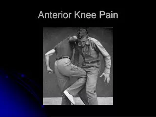
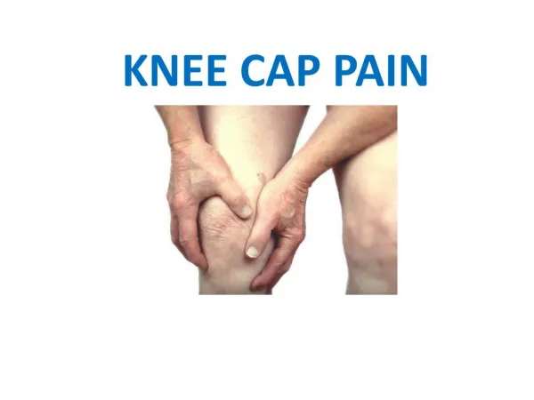
Anterior knee pain • Differential Diagnosis • Injury to quadriceps • Large effusions • Patellofemoral syndrome • Patellar tendonitis • Osgood Schlatter • Inflammatory arthritis (RA, gout, pseudogout) • Septic arthritis http://z.about.com/d/orthopedics/1/0/9/knee.jpg
Lateral knee pain • Least common pain pattern • Differential Diagnosis: • Lateral compartment OA • Lateral collateral ligament injury • Lateral meniscal tears http://www.wbcarrellclinic.com/14x/14xkneemanual/knee_anatomy.jpg
Posterior knee pain • Differential Diagnosis: - Baker’s cyst http://www.dartmouth.edu/~anatomy/assets/surface/knees/surface4.gif
History • History can heighten clinical suspicion but has little value in distinguish between the different types of injuries. • Pain patterns can be broadly categorized into 3 groups: -Symptoms that are most important in arriving at a specific anatomical diagnosis include localized pain, focal swelling, inflammatory change and abnormal noises such as popping or grinding. -Symptoms suggesting the presence of a joint effusion with generalized swelling and impaired bending. -Symptoms reflecting overall change in function including loss of support/collapsing, loss of smooth movement/catching and problems with ambulation.
Physical Exam • The physical exam is a much more sensitive aspect of the visit to make the correct anatomic diagnosis. • It is recommended initiating the exam by focusing on the healthy leg while the patient assumes a position that is most comfortable to them. Examining the healthy knee distracts the patient from the actual maneuvers and creates trust that the examiner is not trying to cause pain. It also serves as the comparison for the affected knee. • Examination consists of inspection, palpation and tests of function.
EXAMINATION MANEUVERS • Right Knee Shown • Lachman test, performed to detect anterior cruciate ligament (ACL) injuries, is conducted with the patient supine and the knee flexed to 20° to 30°. • The anterior drawer test detects ACL injuries and is performed with the patient supine and the knee in 90° of flexion. • The lateral pivot shift test is performed with the patient supine, the hip flexed 45°, and the knee in full extension. Internal rotation is applied to the tibia while the knee is flexed to 40° under a valgus stress (pushing the outside of the knee medially). • The Apley compression test, used to assess meniscal integrity, is performed with the patient prone and the examiner's knee over the patient's posterior thigh. The tibia is externally rotated while a downward compressive force is applied over the tibia. The McMurray test, used to assess meniscal integrity, is performed with the patient supine and the examiner standing on the side of the affected knee. See "Function" section of text for full explanation of all examinations.
Exam • Inspection-Assess gait, muscle atrophy especially in the quadriceps and calves that can occur in ligamental injury, assess for asymmetry that can suggest swelling. Loss of the peripatellar groove on either side of the patella can also be an early sign of effusion. • Palpation-assess for differences in temperature that can suggest inflammation. Assess for effusion with patient supine. Effusion can be detected by ballottement of the patella which is where the excess fluid causes the patella to tap against the femoral condyle when the examiner pushes down on the patella. The tapping sensation is transmitted to the examiner’s fingertips. Crepitus can be noted in joints with cartilage disruption. Joint line tenderness is detected by palpating medial and lateral to the patella in the groove between the femoral condyle and the tibia.
Ligamental Injury • Physical exam is reasonably sensitive in detecting meniscal, ACL and PCL tears 74-81% but less sensitive for detecting other cartilaginous damage 51%. For all lesions except medial meniscus lesions specificity was high 92-96%. • Most common maneuvers include the Lachman test, anterior draw test and lateral pivot shift test. Among these the Lachman maneuver is the most sensitive at 87% [95% CI 0.76-0.98] but its reported specificity is not as high as the pivot test 93% vs 97%[CI 0.93-0.99]. • The Likelihood ratios for the Lachman test are 12.4 +LR and 0.14 –LR. For the anterior drawer test +LR is 3.7 and –LR is 0.6. For the pivot test +LR is 20.3 and –LR is 0.4.
Ligamental Injury • Physical exam is reasonably sensitive in detecting meniscal, ACL and PCL tears 74-81% but less sensitive for detecting other cartilaginous damage 51%. For all lesions except medial meniscus lesions specificity was high 92-96%. • Most common maneuvers include the Lachman test, anterior draw test and lateral pivot shift test. Among these the Lachman maneuver is the most sensitive at 87% [95% CI 0.76-0.98] but its reported specificity is not as high as the pivot test 93% vs 97%[CI 0.93-0.99]. • The Likelihood ratios for the Lachman test are 12.4 +LR and 0.14 –LR. For the anterior drawer test +LR is 3.7 and –LR is 0.6. For the pivot test +LR is 20.3 and –LR is 0.4.
Meniscal injuries • The two specifically studied methods to detect meniscal injuries include the joint line tenderness sign and the McMurray maneuver. • Joint line tenderness is sensitive 76%[CI 0.65-0.87] but not very specific 29% [CI 0.10-0.46] while the McMurray maneuver is specific 97% [CI 0.87-0.99] but not very sensitive 52% [CI 0.35-0.68]. • The +LR for joint line tenderness is 1.1 with a –LR of 0.8. The +LR for McMurray test is 17.3 with a –LR of 0.5.
Meniscal injuries • The two specifically studied methods to detect meniscal injuries include the joint line tenderness sign and the McMurray maneuver. • Joint line tenderness is sensitive 76%[CI 0.65-0.87] but not very specific 29% [CI 0.10-0.46] while the McMurray maneuver is specific 97% [CI 0.87-0.99] but not very sensitive 52% [CI 0.35-0.68]. • The +LR for joint line tenderness is 1.1 with a –LR of 0.8. The +LR for McMurray test is 17.3 with a –LR of 0.5.
Ottawa Rules Rules stated that plain films are indicates in patients with an acute knee injury due to a fall or blow to the knee and have 1 of 4 characteristics: • Age older than 55 • Tenderness at the head of the fibula or isolated to the patella • Inability to bear weight for at least 4 steps i.e. patient can’t take 4 steps • Across the room bearing weight on the injured knee inability to flex the knee to 90 degrees • Most thoroughly validated set of decision rules deciding when to obtain plain films for ruling out fracture. • Resulted from a prospective study of 1047 patients with knee pain for less than 7 days presenting to 1 of 2 Canadian teaching hospital’s ED. Rules have a sensitivity of 100% and specificity of 54% with a Kappa (physician inter-observer agreement) of 0.77. In most studies the use of these rules would have reduced the use of plain knee films by 25%
Osteoarthritis of the Knee • Clinical criteria for OA by the American College of Rheumatology are age older than 50 years, stiffness for less than 30 minutes, crepitus, bony enlargement and no palpable warmth. • If at least 3 of the clinical characteristics are present, the sensitivity for OA is 95% and the specificity is 69%. If 4 criteria are present before the diagnosis of OA the sensitivity decreases to 84% but the specificity increases to 89%.
Radiographic Evaluation- OA • Plain radiograph- May reveal osteophytes, joint space narrowing in medial or lateral compartment. • Osteophytes correlate most with clinical symptoms. Sens and spec for OA 77% and 83%. (Kellgren and Lawrence grading system) • ACR criteria: osteophytes + at least one of the following- age > 50, crepitus, stiffness< 30 min, bony tenderness, enlargement and no warmth. Sens 91% and spec 86% • Sclerosis, cystic degeneration, and meniscus calcification may also be seen. Recommended views include sunrise, weight baring AP and lateral views. • The degree of joint space narrowing over 3 years correlates with future need of TKA in study of 123 patients with 8 year f/u. (Annal Rhem Dis 2005; 64: 1727)
MRI in OA • MRI is not routinely recommended, but is more sensitive for cartilage loss- volume and extent then plain radiographs • May also see bone marrow edema, synovial inflammation • Meniscus and ligament pathology that is more common in severe OA can also be seen on MRI • Quantitative cartilage measurements with MRI, bone marrow edema, meniscus degeneration have been shown to predict progression of OA • Other modalities: • Ultrasound has also been evaluated and has a sensitivity of 76-90% compared to MRI and arthroscopy, a specificity of 92-100% in a recent study with 60 patients. This may be useful in the future, particularly in patients unable to undergo MRI. (Acta Orthop Belg 2006 72:72-6) • CT scan can also show bony changes, but does not show ligament or meniscus injuries well.
MRI in Ligamentous Injury • Recommended prior to arthroscopy • More sensitive, less specific than physical exam findings:
Treatment • Pharmacologic • Tylenol vs. NSAIDs • Injections of Hyaluronic Acid • Glucosamine and Chondroitin • Topical Capsaicin • Steroids • Opiates • Nonpharmacologic
Tylenol vs. NSAIDs • Large crossover trial of diclofenac + misoprostol vs. acetaminophen (Pincus et al. Arthritis Rheum 2001;44:1587-98) • 21 point pain reduction with NSAIDs • 13 point pain reduction with APAP • On this 100 point scale, p<0.001 • But APAP should be 1st line because of toxicity of NSAIDs • Higher doses of NSAIDs are more effective (e.g. ibuprofen 2400 mg/day) • Consider prescribing a PPI or misoprostrol with NSAIDs
Hyaluronic Acid • Data on efficacy are inconsistent • Two metanalyses reported statistically significant but limited efficacy • Evidence of publication bias favoring positive studies • Two large trials showing no efficacy were not published (but summarized in a Cochane Review) NEJM 2006;354:8, p. 844
Glucosamine and Chondroitin • Metanalyses suggests publication bias of early studies • Four recent trials show no efficacy of glucosamine • NIH-sponsored “GAIT” trial (NEJM 2006,354;8:795): • Pain reduction with placebo, glucosamine, chondroitin, or combined glucosamine+chondroitin was similar • Celecoxib (200 mg daily) fared 10% better than placebo (p = 0.008) • However, for the subgroup with moderate to severe pain, the rate of response was better with glucosamine+chondroitin therapy (79%) than with placebo (54%, p = 0.002). • So maybe there is a role for glucosamine/chondroitin in patients with moderate to severe knee OA pain?
Intra-articular Steroids • Demonstrate correct method of knee injection at the: • Knee joint • Prepatellar bursa • Pes anserine bursa • Example: For the knee you need • A 10 mL syringe • An 18 to 22 gauge needle (1.5 in) • 5-7 mL 1% lidocaine or 0.25-0.5% bupivacaine • 2-3 mL betamethasone sodium phosphate and acetate or 2-3 mL methylprednisolone (40 mg/mL) • Draw up the steroid before the lidocaine Effect lasts 1-3 weeks on average
Nonpharmacologic Treatment • Resistance training – in the pool or partial weight-bearing exercises • Unloading with a cane or crutch held contralateral to the affected side • Weight loss • Braces and patellar taping to treat malalignment (but can impair blood return from distal leg) • Acupuncture (one small trial showed a small benefit) • Knee arthroplasty
QUESTIONS • Questions: (Matching 1-6) • 46 yo woman with R> L knee pain, gradually worsening over several weeks, worse with walking down stairs. No catching, no trauma. • 50 yo woman with pain over the medial knee, particularly with crossing her legs. She has pain 2 cm inferior to the medial joint line. • 30 yo man playing soccer, twisting motion of knee with foot planted, developed an effusion, pain and difficulty walking. • 35 yo man with right knee pain, swelling and effusion. No similar prior history. Low grade fevers at home. • 72 yo man with long standing severe OA now with tenderness and swelling in the popliteal fossa. • 67 yo man with h/o DM in the ACC with swollen right knee for 4 days, exquisitely tender with similar problem in right great two last year. • Septic joint b. ACL tear c. Anserine bursitis d. Patellofemoral syndrome • e. Gout f. Baker’s cyst
ANSWERS • D. This is a classic presentation of patellofemoral syndrome which is more common in women and in runners. The lateral compartment of the quadriceps is more developed than the medial, pulling the patella laterally. PE is often benign except for pain with palpation anteriorly over the patella, you may also notice a lateral riding patella. Treat with NSAIDS, strengthening of the medial quads with extension exercises- last 15 degrees of extension. • C. This presentation is suggestive of Pes Anserine bursitis. This can be managed for 6 weeks with NSAIDS, ice. If continued pain, steroid injections at the site of maximal tenderness- insert needle perpendicular to tibia, advance needle to bone, pull back a small amount, aspirate to ensure not in vessel and then inject 40 mg Depomedrol, 1 cc lidocaine. .
ANSWERS • B. The concern here is for a ligamentous injury. If his lower legs was hit from behind while his foot was planted, you might think about ACL tear. Conversely, if the lower leg is struck on the anterior surface, such as in an MVA (against a dashboard) you may be more likely to see a PCL tear. You would look specifically for laxity on the anterior, posterior draw, and Lachman test. An orthopedics consult and likely MRI imaging of the affected knee should be pursued.
ANSWERS • A. This would be concerning for infectious etiology, and you should consider GC. Would ask about pyuria, penile drainage, recent sexual contact. You should do a tap including cultures and gram stain. • F. This is most likely a Baker’s cyst which can be a complication of OA, degenerative changes. Would treat as you treat OA. May give joint injections. Do not inject or aspirate in the popliteal fossa. Ultrasound would confirm. • E. Recurrent monoarticular arthritis- typically limited to 5-10 days, is suggestive of gout. In this patient, making a crystal diagnosis would be helpful. In this scenario, you would need to tap anyway to r/o infectious causes.
QUESTIONS • 54 yo man with blunt trauma to the right knee, acutely painful. There is no effusion or joint laxity and he is able to ambulate. • Would you? A. Get plain films and treat with NSAIDs B. Look for tenderness or patella or fibula and get films only if tender C. Given his age, he needs films even though he can ambulate D. No films now, but if he continues to have pain, order MRI as it is much more sensitive. • Answer is B. Using the Ottawa, there is no indication for plain radiographs for this patient- neg LR 0.11 for fracture. Criteria include age > 55, fibula head tenderness, isolated patella tenderness, inability to flex knee to 90 degrees, inability to bear weight immediately or in ER (4 steps). Manage conservatively with Rest, Ice, Compression, Elevation, NSAIDS. If he continues to have pain at 10 days, plain films should then be obtained
QUESTIONS • 8. 72 yo obese man with bilateral knee pain for several years, increasing pain, decreasing ability to walk, morning stiffness, and crepitus. What is the most likely dx and would you image? • Rheumatoid Arthritis, imaging needed to evaluate for joint damage • Osteoarthritis, imaging needed to evaluate extent of disease • Osteoarthritis, imaging not needed as history and exam consistent with Diagnosis • Parkinson’s with stiffness likely unrelated to knees, refer to neurology
ANSWER 8 C. This presentation is c/w osteoarthritis. Based on clinical predictors alone (see slide 15- sens 95%, spec 69%) which are more sensitive and less specific then clinical plus radiographic findings (sens 91% and spec 86%), the probability that he has OA is 79% based on ACR criteria. There is currently no indication for plain films with initial management. Initial treatment should include APAP, then NSAIDS, resistance training, weight loss. If the above treatments are not effective, may then proceed to intra-articular steroids, consider arthroplasty.
72 yo man with long standing severe OA now with tenderness and swelling in the popliteal fossa. Your next step is A. Drain popliteal fossa B. Aspirate joint C. Rest, Ice, Compression, Elevation D. Xray and refer to ortho Answer 9: B This is most likely a Baker’s cyst which can be a complication of OA, degenerative changes. Would treat as you treat OA. May give joint injections. Do not inject or aspirate in the popliteal fossa. Ultrasound would confirm
Reference • Anderson RJ. Evaluation of the adult patient with knee pain. www.uptodate.com search phrase: knee pain. • Calmbach WL, et al. Evaluation of Patients Presenting with Knee Pain: Part I. History, Physical Examination, Radiographs and Laboratory Tests. American Family Physician 2003; 68(5): 907 – 912. • Heintjes EM, et al. Knee disorders in primary care: design and patient selection of the HONEUR knee cohort. BMC Musculoskelet Disord 2005; 6: 45 – 55. • Jackson JL, et al. Evaluation of Acute Knee Pain in Primary Care. Annals of Internal Medicine 2003; 139: 575 – 588. • Solomon DH, et al. Does This Patient Have a Torn Meniscus or Ligament of the Knee? Value of the Physical Examination. JAMA 2001; 286 (13): 1610 – 1620. • Evaluating the Patient with a Knee Injury Family Practice Management 3/05 67-70 • Up to Date Version 14.1