Evidence for Precursor-Product Relationship in RNA Processing
700 likes | 798 Vues
This recap summarizes key experiments demonstrating the precursor-product relationship in eukaryotic RNA processing, highlighting observations and experiments that led to this understanding. It discusses RNA stability, self-splicing introns, and the complex machinery involved in splicing. The role of snRNPs, spliceosome assembly, and splicing mechanisms are explained in detail, showcasing the intricacies of RNA processing in eukaryotes.

Evidence for Precursor-Product Relationship in RNA Processing
E N D
Presentation Transcript
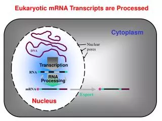
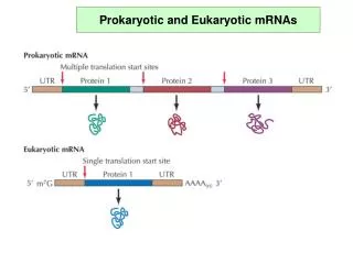
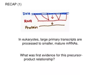
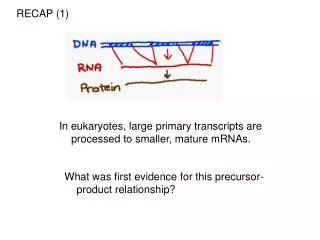
RECAP (1) In eukaryotes, large primary transcripts are processed to smaller, mature mRNAs. What was first evidence for this precursor-product relationship?
Observation: Nuclear RNA pool consists of very high molecular weight species as well as lower molecular weight. Darnell asked if there is a relationship between the high and low molecular weight RNAs DNA
Experiment: Treat cells with UV for varying periods of time. Thymidine dimers will form, blocking transcription. To assess the effects on the two pools of RNA, pulse cells with 3H-Uridine and measure counts in each pool DNA Example UV dose that hits 1X/1000 bp X X X X X X X X X X If long RNAs are precursors then both long and short pools should exhibit comparable UV sensitivity If long and short RNAs are independently transcribed, then they should exhibit different UV sensitivity
Experiment: Treat cells with UV for varying periods of time. Thymidine dimers will form, blocking transcription. To assess the effects on the two pools of RNA, pulse cells with 3H-Uridine and measure counts in each pool DNA Example UV dose that hits 1X/1000 bp X X X X X X X X X X If long RNAs are precursors then both long and short pools should exhibit comparable UV sensitivity If long and short RNAs are independently transcribed, then they should exhibit different UV sensitivity
RECAP (2) RNA is unstable – it can cleave itself. RNA can fold into complex 3D structures. Self-splicing introns utilize this suicidal tendency and contortionist ability to direct self-cleavage at precisely defined sites
RECAP (3) Splicing in eukaryotes probably relies on the same chemistry as self-splicing group II introns. Splicing substrates in eukaryotes much more varied, and can’t rely on 2o structure alone to define splice sites. A complex RNA+protein machine is used to precisely define splice sites.
The spliceosome is made up of 5 small nuclear ribonucleoprotein subunits + > 100 proteins. These snRNPs are called: U1, U2, U4, U5, U6, and assemble in a stepwise pathway onto each intron. There are also many additional non-snRNP proteins in the spliceosome.
Structures of the Spliceosomal snRNAs U1, U2, U4, U5 RNA Pol II transcripts TriMethyl G Cap Bound by Sm Proteins U6 RNA Pol III transcript Unusual Cap Not bound by Sm proteins Each snRNA has a specific sequence and secondary structure and is bound by additional specific proteins
The earliest snRNP to bind to the pre-mRNA is U1, which uses its snRNA to base-pair to the 5’ splice site.
The U2 snRNP binds to the branchpoint via RNA/RNA base-pairs to create a bulged A residue. This forms the pre-spliceosomal “A” complex.
The protein U2AF (U2 Auxiliary Factor) binds to the Polypyrimidine tract and the AG of the 3’ splice site and helps U2 snRNP to bind to the branchpoint . U2AF65 35
U2AF Splice sites do not always perfectly match the consensus sequences. Thus, the base-pairing interactions between the snRNAs and the pre-mRNA are not always the same. Pre-spliceosome
Pre-mRNA The interactions of U1 with the 5’ splice site and U2 with the branchpoint were proven by creating mutant splice sites that bound the snRNA so poorly that splicing was inhibited. Compensating mutations in the snRNA that restored complementarity (base-pairing) with the splice site restored splicing.
The full spliceosome is formed from the pre-spliceosome by the addition of the U4/U5/U6 Tri-snRNP.
Cap 5’ U6 snRNA U4 snRNA In the U4/U6 Di-snRNP and the U4/U5/U6 Tri-snRNP, the U4 and U6 snRNAs are base-paired to each other. This interaction is later disrupted in the formation of the active spliceosome.
After the formation of the full spliceosome, the U1 and the U4 snRNPs are detached and the remaining U2, U5 and U6 snRNAs are rearranged. This conformational change creates the catalytic spliceosome.
U6 snRNA U2 snRNA intron U5 snRNA In the catalytically active spliceosome, the U2, U5 and U6 snRNAs make very specific contacts with the splice sites.
The two transesterification reactions of splicing take place in the mature spliceosome.
After the second transesterification reaction, the spliceosome comes apart. The snRNPs are recycled, and the spliced exons and the lariat intron are released.
The lariat intron is debranched by Debranching Enzyme returning it to a typical linear state. This linear intron is quickly degraded by ribonucleases.
Mobile genetic elements provide an example of RNP complexes in which proteins and RNAs cooperate for specificity group II self-splicing intron encodes an endonuclease (E) maturase (M) and reverse transcriptase (RT) that are used for integration of the mobile element back into the genome. The intron, E, M, and RT form an RNP and the 2’OH of the intron directs cleavage of the first strand of the target DNA. Group II self-splicing intron forms the core of an RNP that can direct cleavage of other nucleic acid polymers.
U6 snRNA U2 snRNA intron U5 snRNA In the catalytically active spliceosome, the U2, U5 and U6 snRNAs make very specific contacts with the splice sites. What are the proteins doing in catalysis?
A tale of the U5 protein, Prp8. Prp8 mutants are splicing defective. Many Prp8 mutations suppress splicing defects caused by 5’-SS, 3’-SS and branch point mutations. Prp8 cross links to crucial U5, U6, 5’-SS, 3’-SS and branch point residues. Prp8 interacts with Brr2 and Snu114, which unwind U4/U6 and allow U2 to pair with U6
Crystal structure of Prp8 reveals a cavity of appropriate dimensions to position spliceosomal RNAs for catalysis. Group II intron Prp8 Structural domains of Prp8 (endonuclease, reverse transcriptase) suggest ancient evolutionary origins as a homing endonuclease.
Splicing is dynamic, with sequential regulated alterations in RNA:RNA and RNA:protein interactions
Splicing error rates range from 1 in 1000 to 1 in 100,000 DEAD-box RNA helicases implicated in quality control
Transitions regulated by DEAD-box ATPases • Monomeric (vs. “AAA” ATPases) • RNA-dependent ATPases • ~300 aa domain with 7 signature motifs (e.g. eponymous tetrapeptide) • 2 RecA-like folds bind ATP, RNA (“closed form”) • Conformation opens upon ATP hydrolysis (i.e. switch-like) • 8 essentialspliceosomal DEAD-box ATPases in yeast (more in mammals) • In vitro: • Most catalyze RNA-dependent ATP hydrolysis (ATPase) • Some catalyze ATP-dependent RNA unwinding (“helicase”) • In vivo???? • Likely most are “RNPases”, destabilizing RNA:protein complexes
The story of one helicase: PRP16 Prp16 is required for the second chemical step: - Immunodeplete Prp16, inc. extract w ATP, P-32 substrate -> LI - Now deplete ATP, then add back rPrp16 + ATP -> Exon ligation - Instead, add back rPrp16 – ATP -> No splicing, but Prp16 bound Conclude: Prp16 can bind to LI but requires ATP hydrolysis for release and promotion of the second chemical step
The story of one helicase: PRP16 • Prp16-1 mutant was identified in a screen for reduced-fidelity mutants: • Mutate branchpoint A to C in a splicing reporter • Mutagenize cells ->Select for improved splicing of reporter • Repeat selection by mutagenesis of cloned PRP16 gene -> • - New suppressors all map to the conserved DEAD-box domain • In vitro, mutant Prp16 proteins have reduced ATPase activity • Conclude: • Prp16 modulates the fidelity of splicing by an ATP-dependent mechanism
The story of one helicase: PRP16 Hypothesis: Prp16 promotes fidelity 1) branchpoint mutations -> slow conformational rearrangement -> rejection 2) suppressor mutations in Prp16 -> more time
The story of one helicase: PRP16 How to discriminate between “correct” vs. “incorrect”? A “slow” spliceosome -> ATP-dependent rejection of WT substrate. Conclusion: ATPases promote specificity by discriminating against slow substrates
PRP16: functions at 2 steps PRP16 binds before 5’ss cleavage and acts as a sensor to promote discard of suboptimal substrates PRP16 promotes exon-exon ligation
Questions How are the intervening sequences removed? How are the splice sites identified?
How are the splice sites identified? In higher eukaryotes, there isn’t much sequence information encoded in the 3’ss, 5’ss, or branch point
How are the splice sites identified? Minor spliceosome, consists of U11, U12, U4atac, U6atac, and U5 About 100-fold less abundant than major spliceosome Splices ~ 0.2% of introns in vertebrates
How are the splice sites identified? Human Dystrophin gene 260 kb intron 2.4 Mb Genes in higher eukaryotes have many exons and introns can be very large
How are the splice sites identified? The same primary transcript can be spliced many different ways (estimated 90% of genes experience alternative splicing)
How are the splice sites identified? Because of the intron length and lack of specificity of splice sites, most introns contain numerous cryptic splice sites in addition to bona fide alternative splice sites.
How are the splice sites identified? outcomes of 5’ ss mutants x 1. activates cryptic 5’ ss, but only if there is one within 100-300 bp of original 5’ ss x 2. skip the exon altogether and ignore perfectly good 3’ and 5’ ss of the upstream intron
How are the splice sites identified? beta-globin mutants that create a new 3’ ss within an intron: x also create a new exon???
In multicellular organisms, exons are recognized as units prior to assembly of the spliceosome across the long introns. This “exon definition” step involves interactions between the splice sites across the exon and special sequences in the exon called Exonic Splicing Enhancers (ESE). The sequences in exons are selected to not just code for particular peptide sequences, but also for binding of regulatory proteins to ESE’s.
How are the splice sites identified? Boundaries between introns & exons are recognized through its interaction with multiple proteins either across exon or intron Intron definition: Uses intron as the unit of recognition mechanism. Complex forms through stabilized protein interactions across the intron SR SR SR RS 70K U2AF35 RS U2AF RS SF2 Exon 1 Exon 2 A SF1 U1 snRNP Intron Definition SR Exon Definition: Complex can easily form stabilized protein interactions across the exon. Excises out the flanking introns SR RS 70K SR U2AF35 RS U2AF RS SF2 Exon A SF1 U1 snRNP Exon Definition (Cote, Univ. of Ottawa) Stable interaction confirms accuracy of splice site choice
Why are exons preferentially recognized? • Differential size distributions of exons (~50 to 300 nt) vs. introns (<100-100,000 nt) • SR protein - preferentially binds to exon sequences • - mark the 5’ & 3’ splicing sites in conjunction w/ U1 & U2 during transcription • hnRNP - heterogenous nuclear ribonucleoproteins (twice the diameter of nucleosome) • - consists at least eight different proteins • - compacts introns, thereby masking cryptic splicing sites • - preferentially binds to introns, but also bind to exons, although less frequently
Cross-exonbridging interactions involve SR domains of U2AF, U170K • And 1 or more SR-family proteins • ~12 in mammals (and # AS isoforms!) • Tissue-specific differences in concentration • RRMs vary in degree of sequence preferences • Outstanding question: • What triggers the switch from Exon- to Intron-Defined interactions?
Splicing is co-transcriptional and all introns assayed are spliced within 5-10 minutes of transcription of the downstream exon and 3’ splice site, regardless of intron size (1 kb or 240 kb)
Defining an exon involves the specific stabilization or destabilization of splice site recognition • Stabilization: exon inclusion • Destabilization: exon skipping
Regulation of alternative splicing involves the specific stabilization or destabilization of splice site recognition • Stabilization: exon inclusion • Destabilization: exon skipping
How would you identify cis-regulatory sequences responsible for alternative splicing ? Mutational analysis finds an element necessary for exon inclusion Transfection Reporter Plasmid Alternatively spliced Not alternatively spliced Examine RNA Splicing of Transfected Splicing Reporters to identify cis-regulatory regions