Download
1 / 65
740 likes | 1.2k Vues
Nucleotides and Nucleic Acids. Dr. W. McLaughlin. Learning Objectives:. The types of nucleic acids The structure and components of nucleic acids The conformations of DNA The base composition of DNA Function of nucleic acids. Why learn about Nucleic Acids?.

E N D
Nucleotides and Nucleic Acids Dr. W. McLaughlin
Learning Objectives: • The types of nucleic acids • The structure and components of nucleic acids • The conformations of DNA • The base composition of DNA • Function of nucleic acids
Why learn about Nucleic Acids? • Knowledge of the anatomy of the gene is as important in medical practice as knowledge of the anatomy of the heart
There have been numerous advances in the areas of molecular biology and biotechnology in medicine but the biggest could be the Human Genome Project • The information, it is hoped will revolutionize the detection, prevention and treatment of conditions from cancer to depression to old age itself
The Future Medical Doctors… …will drip droplets of our genes onto a biochip to figure out if we have the kind of prostrate cancer that will kill or not, or to figure out if ours is the kind of leukaemia that respond to this drug rather than that one …medical records will include the complete genome ..treat patients as a biochemical and genetic individual
THE STRUCTURE OF NUCLEIC ACIDS Both DNA and RNA are known as nucleic acids • Nucleic acids are high-molecular-weight polymeric compounds • On complete hydrolysis yields pyrimidine and purine bases, a sugar component and phosphoric acid. • Partial hydrolysis yields compounds known as nucleosides and nucleotides
Structure of Nucleotide • A nucleotide consists of three things: • A nitrogenous base, which can be either adenine, guanine, cytosine, or thymine (in the case of RNA, thymine is replaced by uracil). • A five-carbon sugar or pentose sugar • One or more phosphate groups
Cyclic 5-carbon sugar • D-ribose in RNA • 2'-deoxyribose in DNA
Purine Bases • Adenine & Guanine • Other naturally occurring derivatives • Hypoxanthine • Xanthine • Uric acid
Pyrimidine Bases • Thymine, cytosine and uracil • 5-methylcytosine • 5-hydroxymethylcytosine. • N4-methylcytosine • N6-methylcytosine
Phosphate group • a molecule of Phosphoric acid, PO43
Nucleosides • Purine or a Pyrimidine base + ribose or deoxyribose = Nucleoside. • ribose = ribonucleosides • 2’-deoxyribose = deoxyribonucleosides
Nucleotides • Purine or a pyrimidine linked to ribose or deoxyribose and phosphoric acid = Nucleotide • ribose = ribonucleotides • 2’-deoxyribose = deoxyribonucleotides
Nucleic Acids • Nitrogenous base attached to the sugar by N-glycosidic bonds to carbon # 1 of the sugar • Sugar is attached at position N-1 of the pyrimidine base • Sugar is attached at position N-9 of the purine base • Phosphate forms a bond with the sugar through phosphodiester linkages between 5' –OH of the sugar and the negatively charged oxygen group
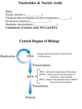
Primary structure of nucleic acids Nucleotides join through phosphodiester linkages between 5’ and 3’ carbon atoms to form nucleic acids When many nucleotides subunits combine, the result is the large single-stranded polynucleotide or Nucleic Acid
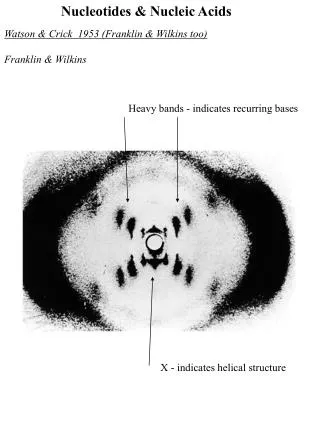
Secondary structure of DNA • DNA exists as a double helix • In April 1953, Watson and Crick proposed the structure of DNA
Secondary structure of DNA • two complementary polymeric chains forming a regular right-handed double helix • the two stands run in opposite directions (antiparallel alpha-helices), and are of opposite polarity • the rails of the ladder runs in opposite direction and contain alternating units of deoxyribose sugar and phosphate.
2° structure of DNA • the sugar and phosphate groups are always linked together by 3’ - 5’ phosphodiester linkages. • the purine and pyrimidine bases are flat (planar), are relatively water-insoluble and are stacked tightly on top of one another like a pile of plates, forming the steps of the helical ladder. • the bases are arranged at right angles to the long axis of the polynucleotide chain.
2° structure of DNA • each step is composed of a pair of nucleotide- a base pair held together by weak hydrogen bonds. • the order of the purine and pyrimidine bases along the chain is highly irregular, varying from one molecule to the other. • the chain is not straight but is wound helically around a central axis, one full turn ( the pitch) of the helix extending 3.4 nm (34 Å), and there are 10 bases per turn
2° structure of DNA • the bases are separated by a spacing of 0.34 nm (3.4 Å). • the width of the double helix is 2 nm (20 Å). • the chains are complementary, the sequence of bases on one strand is the exact complement of the other strand. • Adenine always pair with thymine and cytosine always pair with guanine.
Hydrogen bonds • Essential for 3-D structure of DNA • Not the main contributor of DNA stability • Important for the specificity of the helix • Allow for only complementary strands to pair • A=T; G=C • Complementary nature allow DNA to carry information
Bases pair in a Specific way • Purines are larger structures than pyrimidine, if two purine are paired their dimensions are too great to fit the constant diameter of the double helix (2 nm) while the dimensions of the two pyrimidine are too small • The specificity of position of the H atoms that can participate in bonding. It is essential that the hydrogen bonds have relatively stable positions to have the biological functioning of DNA.
Tautomeric Shifts • H-atoms do undergo shift to other positions to form new pairing interactions • the nitrogen atoms attached to the purine and pyrimidine rings are usually in the amino (NH2) form and only rarely assume the imino (NH) configuration • the oxygen atoms attached to C6 atoms of guanine and thymine normally have the keto (C-O) form and rarely take up the enol (COH) configuration.
Tautomeric Shifts • if the H-atom normally present at the 6-amino position in adenine shifts to the N1 position, Adenine will pair with Cytosine instead of with Thymine
Base composition of DNA • Chargaff described fundamental features of DNA as revealed by chemical analysis. • Chargaff was the first to draw attention to certain regularities in the composition of DNA • Chargaff’s Rule is true because of the strict H-bond forming rules
Chargaff’s Rules • purines = pyrimidines • amino bases (A & C) = keto bases (G & T) • between the amounts of A & T, and between the amounts of G and C, ( A = T and G = C). • Wide variations in the molar proportions of bases although DNA from different organs and tissues of any one species are essentially the same. • A+T/G+C, (base ratio) may vary widely between species, and remains constant for any one species.
Molar Proportion of Bases in DNA from various sources ____________________________________________ Source A G C T A+T/G+C _____________________________________________ Bovine thymus 28.2 21.5 21.2 27.8 1.3 Bovine spleen 27.9 22.7 20.8 27.3 1.3 Bovine sperm 28.7 22.2 20.7 27.2 1.3 Wheat germ 27.3 22.716.8 27.1 - Yeast 31.2 18.7 17.1 32.9 1.8 E. coli 26.0 24.9 25.2 23.9 1.0 E. aerogenes 21.0 28.0 29.0 22.0 0.7 C. perifringens 34.0 15.0 16.0 35.0 2.2 M. tubercolosis 15.1 34.9 35.4 14.6 0.4 X174 24.3 24.5 18.2 32.3 - _____________________________________________
DNA Conformations • The major forms of DNA are: • the B-form, basically describes the Watson and Crick model • the A-form DNA
B-form DNA • Represent the conformation of most DNA found in cells • Exists under most physiological conditions • The main features are: the pitch, the angle of tilt, the distinct major and minor grooves • Pitch 3.4 nm • 10 bases/full turn • Long and thin
Major and Minor Grooves • Two asymmetrical groves • Larger = Major; Minor = Minor • Arise because of the geometrical configurations of the bonds • The groves expose the bases • Recognised by proteins
A-form DNA • The base pairs tilt some 30 so that successive base pairs occur every 0.28 nm • Adopted by DNA under low humidity • 11 bases/full turn • Is short and broad and has deeper and narrow major grooves • he 2’-OH of ribose prevents RNA from forming the classic B-helix
Z-form DNA • Alternating purine and pyrimidine residues (dinucleotides CGCGCGCGCGC) can fold up into left-handed as well as right handed helices. • One deep helical groove
50 Yrs after the discovery of DNA • Deciphering of the human genome • Genetic engineering of animals and crops • Use of gene therapy to treat human diseases • Designing better drugs • Admissibility in courts in criminal cases • DNA chips
Properties of DNA • Denaturation & Renaturation of DNA The DNA double helix can unwind to form single strands when subjected to : • extremes of pH • increased temperature • decreased dielectric constant by alcohols, ketone, etc. • exposure to amides or urea
DNA denaturation • denature when the DNA changes from a double helix to a random coil • melting or transition temperature Tm • measured at 260 nm • hyperchromic effect
Denaturation of DNA • The nature of the melting transition is affected by 3 factors: • The G+C content of the DNA • The nature of the solvent. • The nature of the DNA.
G+C Content • DNA with higher G+C is more stable and have a higher melting temperature • GΞC • A=T