Download
1 / 61
640 likes | 933 Vues
BIO 169 HUMAN DEVELOPMENT AND INHERITANCE CHAPTER 29. created by Dr. C. Morgan. TOPICS. Introduction and Overview Fertilization The First Trimester The Second and Third Trimesters Labor and Delivery Postnatal Development Genetics, Development, and Inheritance.

E N D
BIO 169 HUMAN DEVELOPMENT AND INHERITANCE CHAPTER 29 created by Dr. C. Morgan
TOPICS Introduction and Overview Fertilization The First Trimester The Second and Third Trimesters Labor and Delivery Postnatal Development Genetics, Development, and Inheritance
Introduction and Overview Objectives Define development. Review the process of differentiation. Define fertilization. Define growth. Distinguish between embryonic and fetal development. Distinguish between prenatal and postnatal development. Define inheritance.
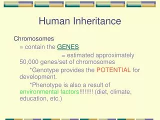
Introduction and Overview Development is the gradual modification of a fertilized ovum into the functional anatomical structures present at maturity. Differentiation is the selective activation of genes leading to cell populations that have distinct functions and form distinct tissue types that comprise the various organs and organ systems. Developmentinvolves generations of cell divisions and differentiation along with establishing new relationships of cells that form tissues. Chemical signals induce the developmental changes resulting from new relationships among cells and tissues Growth, an increase in size, is distinct from development.
Introduction and Overview (cont) Fertilization is the fusion of the genetic material of two haploid gametes to produce a diploid cell. Human embryological development is the time span from fertilization through 2 months of gestation. Fetal development is the time span from the beginning of the third month until birth. Prenatal development is the time span prior to birth. Postnatal development is the time span from birth until maturity. Inheritance is the transfer of genetic information from generation to generation.
TOPICS Introduction and Overview Fertilization The First Trimester The Second and Third Trimesters Labor and Delivery Postnatal Development Genetics, Development, and Inheritance
Fertilization Objectives Describe what a sperm cell must do in order to fertilize an ovum. Discuss the functional roles of the ovum and sperm. Learn what is meant by capacitation. Discuss the fate of sperm present in the ejaculate. Using an illustration, describe the oocyte changes with fertilization. Preview prenatal development.
Fertilization An ovulated ovum is surrounded by its plasma membrane and up to several layers of follicular cells (the coronaradiata) which establish a barrier to sperm penetration. Thus, sperm must secrete digestive enzymes that weaken the corona radiata to create an opening where the sperm cell is able to enter the ovum cytoplasm. Since the only contribution of the sperm is its haploid set of chromosomes, all the organelles, cytosol, metabolic machinery, and nutrients along with the other haploid genome are provided by the ovum. The cytoplasmic rich egg is 2000 times larger than a sperm. Although sperm are motile when introduced into the vagina, they must undergo some biochemical changes (capacitation) before they are capable of fertilization.
Fertilization (cont) Of the approximately 100–200 million sperm in an ejaculate, only about 100 survive to make it to the ovum surface. The collective enzymes present in the acrosome of these sperm are needed to weaken the corona radiata. Only one sperm cell is permitted to fertilize an egg because as soon as it penetrates the cell membrane, a chemical reaction takes place in the zona pellucida to exclude additional sperm. Oocyte activation follows sperm entry as metabolic activities of the ovum increase in preparation for the cleavage cell divisions. Fig. 1 a
Fertilization (cont) Fig. 1 b
Fertilization (cont) Fertilization is the first event of prenatal development also called pregnancy or gestation. For purposes of discussion, pregnancy is considered in three month intervals or trimesters. First trimester– embryonic and early fetal development Second trimester– organs and organ systems complete development Third trimester– rapid fetal growth and establishment of full functionality in organ systems
TOPICS Introduction and Overview Fertilization The First Trimester The Second and Third Trimesters Labor and Delivery Postnatal Development Genetics, Development, and Inheritance
The First Trimester Objectives Using an illustration, describe cleavage and blastocyst formation. Using an illustration, describe the stages of the implantation process. Discuss the processes that lead to the germ layers. Describe the formation and purpose of the extraembryonic membranes. Discuss the establishment of the placenta. Illustrate the events during early embryogenesis.
The First Trimester cleavage implantation placentation embryogenesis Cleavage and blastocyst formation Implantation in uterine wall Fig. 2
The First Trimester (cont) There are several generations of cleavage divisions that occur every 10 – 12 hours after fertilization. After 5 days, there is a hollow ball called a blastocyst that contains an inner cell mass surrounded by cells that make up the trophoblast. The inner cell mass forms the embryo. By day 6, the blastocyst is in the uterus and its cells are able to obtain some nutrients from the fluid secreted by the glands of the endometrial functional zone. By day 8, the trophoblast cells contact the endometrium, the initial step in implantation. The surface trophoblast cells divide rapidly with some losing their cell membranes to create a multinucleate layer of cytoplasm called the syncytial trophoblast.
The First Trimester (cont) The cellular trophoblast remains intact and in contact with cells of the inner cell mass. The syncytial trophoblast digests its way into the endometrium until the entire structure is buried within the endometrium. Implantation continues as the trophoblast digests more uterine tissue and grows around uterine capillaries, destroying capillary walls allowing maternal blood to flow through channels (lacunae) in the trophoblast. 16 Fig. 3
The First Trimester (cont) Villi extend further into the endometrium. The inner cell mass separates from the trophoblast to create the amniotic cavity. Inner cell mass forms the 2 layered blastodisc. Fig. 4 17
The First Trimester (cont) Gastrulation forms three “germ layers” TABLE 1: germ layer fates cells migrate Fig. 4
The First Trimester (cont) selected germ layer derivatives (TABLE 1) Ectoderm: integumentary system nervous system pituitary gland, adrenal medullae mucous epithelium of nasal passages, mouth, anus salivary glands Mesoderm: skeletal system muscular system cardiovascular system urinary system; adrenal cortex lymphatic system gonads and ducts all connective tissue linings Endoderm:mucous epithelium of digestive tract, liver, pancreas, urinary bladder, gamete stem cells and distal reproductive ducts, thymus and thyroid glands
The First Trimester (cont) All organs systems are derived from cells of the germ layers. Outside the embryo are four extraembryonicmembranes which are also derived from the germ layers. *Mesodermal cells migrate around the endodermal pouch under the blastodisc to form the yolk sac (site of early blood cell formation; becomes part of umbilical cord). *Mesodermal cells migrate around the inner cellular trophoblast to form the chorion (forms placental tissue). *Mesodermal cells migrate around the outside of the amnionic cavity to form the amnion (forms a cavity containing a protective fluid that surrounds embryo). *An endodermal extension surrounds the mesoderm to form the allantois (base becomes part of bladder and remainder part of the umbilical cord).
The First Trimester (cont) nutrient source TABLE 2: Summary of Prenatal Development Fig. 5 a
The First Trimester (cont) Fig. 5 b
The First Trimester (cont) Chorionic villi with embryonic blood vessels extend further into endometrium placentation Fig. 5 c
The First Trimester (cont) nutrients are obtained by transport from maternal blood in the placenta into the embryonic vasculature Fig. 5 d
The First Trimester (cont) Paired umbilical arteries and one vein are part of the umbilical cord Fig. 5 e
The First Trimester (cont) Placental structure active and passive transport uterus with embryo removed Fig. 6
The First Trimester (cont) The placenta is also an endocrine organ with hormones secreted by the syncytial trophoblast cells into the maternal blood. Human chorionic gonadotropin (hCG) appears soon after implantation (basis for pregnancy test) with the role of maintaining the corpus luteum. Human placental lactogen (hPL) and placental prolactin target mammary glands. Relaxin, from the placenta and corpus luteum, causes the pubic symphysis to become flexible, dilates the cervix, and suppresses oxytocin secretion to delay labor. Placental progesterone maintains the endometrium. Placental estrogen (3rd trimester) helps trigger labor.
The First Trimester (cont) Embryogenesis vertebraemuscles Fig. 8 a
The First Trimester (cont) fiber-optic camera image Fig. 8 b
The First Trimester (cont) fiber-optic camera image Fig. 8 c
The First Trimester (cont) Table 2 Fig. 8 d
TOPICS Introduction and Overview Fertilization The First Trimester The Second and Third Trimesters Labor and Delivery Postnatal Development Genetics, Development, and Inheritance
The Second and Third Trimesters Objectives Characterize the fetal changes during the second and third trimesters. Discuss the major changes involving maternal physiology. Describe the structural and functional changes in the uterus including hormonal interactions.
The Second and Third Trimesters The rudiments of all organ systems are established in the first trimester so the second and third trimesters are a time of growth and establishment of physiological function. The maternal organ systems must meet all the nutritional, respiratory, and waste disposal needs of the fetus. To do this, maternal systems must make far reaching adjustments which include the following: *increased respiratory rate and tidal volume *increased blood volume *increased nutritional and vitamin requirements *up to a 50% increase in filtration through the kidneys *increase in both the uterine and mammary gland size
The Second and Third Trimesters 16 week fetus Fig. 10 a
The Second and Third Trimesters displacement of visceral organs by fetus Fig. 10 c
The Second and Third Trimesters (cont) Placental progesterone inhibits contraction of the uterine smooth muscle until overpowered by estrogens. The placental estrogens increase the sensitivity of the uterine smooth muscle. Near the end of pregnancy, estrogen production increases. The sensitive myometrium is stimulated to contract by rising levels of oxytocin released from the posterior pituitary. Oxytocin release is triggered by circulating estrogens and activated stretch receptors in the cervix. Late in pregnancy, uterine tissue produces prostaglandins which also help trigger uterine contractions. Late in pregnancy, the fetus releases additional fetal oxytocin which probably initiates labor and delivery.
The Second and Third Trimesters (cont) Initiation of labor Relaxin acts on pubic symphysis and helps dilation Fig. 11
TOPICS Introduction and Overview Fertilization The First Trimester The Second and Third Trimesters Labor and Delivery Postnatal Development Genetics, Development, and Inheritance
Labor and Delivery Objectives Describe the events of the dilation stage. Describe the events of the expulsion stage. Describe the events of the placental stage. Discuss premature labor. Discuss multiple births.
Labor and Deliver The expulsion of the fetus is called parturition. Smooth muscle contractions of the myometrium begin at the top of the uterus and proceed toward the cervix. A positive feedback cycle involving oxytocin causes the contractions to become progressively stronger and more frequent to move the fetus toward the cervical canal. Labor proceeds in three stages: dilation, expulsion, and placental. *Contractions that last 30 seconds at 10 to 30 minute intervals cause dilation of the cervix. It usually take 8 hours or more to complete dilation. The amnion ruptures near the end of the dilation stage.
Labor and Deliver (cont) Fully Developed Fetus Fig. 12
Labor and Deliver (cont) Dilation Stage Fig. 12 a
Labor and Deliver (cont) *Expulsion begins when the fetus has forced the cervix completely open and begins to pass through the vagina. Typically expulsion takes less than 2 hours. If the vaginal opening is small, a cut through the perineal musculature (episiotomy) is made between the vaginal and anal openings to prevent tissue tearing. Delivery is arrival of the fetus into the outside world. If complications are encountered or anticipated, a cesarean section surgical procedure can be performed to remove the fetus through an abdominal incision. *Due to the shrinking uterus, the placenta is dislodged and ejected within an hour along with substantial amounts of maternal blood (the placental stage).
Labor and Deliver (cont) Placental Stage Fig. 12 c
Labor and Deliver (cont) Premature labor occurs prior to completion of normal fetal development. Survival of premature infants is related to their body weight. Those weighing less than 400 g (14 oz) have little chance of survival while those weighing between 500 g and 1 kg have a 50% chance of survival (often with abnormalities). Those weighing more than 1 kg have a very good chance of survival. With the advent of fertility drugs, in vitro fertilization, and other mechanisms of manipulating the reproductive process, multiple births are on the rise. Dizygotic twins are from separate eggs and sperm. Identical twins result from separation of early cleavage cells
TOPICS Introduction and Overview Fertilization The First Trimester The Second and Third Trimesters Labor and Delivery Postnatal Development Genetics, Development, and Inheritance
Postnatal Development Objectives List the postnatal developmental stages. Discuss lactation. Postnatal Development The postnatal developmental life stages are– neonatal, infancy, childhood, adolescence, and maturity. *The neonatal period is from birth to one month thereafter. Neonates have heart rates around 120 –140 and respiratory rates around 30 bpm. Neonates who are breast fed first get a glandular secretion called colostrum which is high in protein (many are antibodies) and gradually milk after 2 to 3 days.
Postnatal Development (cont) Milk let-down reflex milk composition oxytocin triggers contraction of myoepithelial cells water proteins amino acids lipids sugars salts antibodies 750 C / liter Human placental lactogen and placental prolactin simulate mammary glands during gestation Fig. 13
Postnatal Development (cont) The most rapid growth is during fetal development and thereafter it gradually slows. Growth hormone, adrenal steroid hormones, and thyroid hormones control growth. Growth does not occur uniformly in all cells and organ systems which results in a change in proportions over time. 50 Fig. 14