Download
1 / 48
600 likes | 2.78k Vues
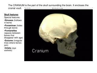
The CRANIUM is the part of the skull surrounding the brain. It encloses the cranial vault. Skull features Special features: Sinuses (hollows in bone) Foraminae (holes through bone) Fontanelles (spaces between bones that disappear with age) Sutures (irregular lines where bones join)

E N D
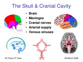
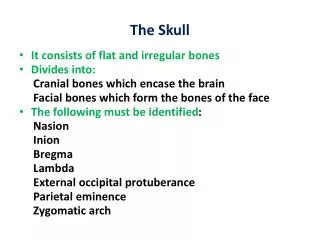
The CRANIUM is the part of the skull surrounding the brain. It encloses the cranial vault. • Skull features • Special features: • Sinuses (hollows in bone) • Foraminae (holes through bone) • Fontanelles (spaces between bones that disappear with age) • Sutures (irregular lines where bones join) • Orbits (eye sockets)
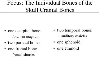
Bones of the cranial vaultTwo frontal, two parietal, one occipital, two temporal, one sphenoid (8 bones) Frontal Parietal Occipital Temporal Sphenoid
Skull sutures Coronal Squamous Lamdoid Sagittal
Skull landmarkswhere sutures meet N.B. Remember the pterion! It is the weak spot above the middle meningeal artery. Bregma Pterion Asterion TMJ Lambda Mastoid process
Development of the skull Anterior Fontanelle (Bregma) F P S T Posterior Fontanelle (lamda) Anterolateral Fontanelle (pterion) Posterolateral Fontanelle (asterion)
The occipital bone overlies the occipital cortex and the cerebellum
The foramen magnum is where the spinal cord exits the skull. Note the occipital condyles on each side. This is where the skull rests and articulates on the atlas condyle
Note that the occipital bone projects surprisingly far forward at the base of the skull. It articulates with the sphenoid bone. Anterior-most part of occipital bone slopes steeply upwards. This is the ‘clivus’
The parietal bone overlies the parietal cortex. Its anterior end is part of the pterion
The temporal bone overlies the temporal lobe. It is a complex bone with several foramina for nerves in the petrous (‘pete-russ’) part
External auditory meatus Temporomandibular joint
You can feel the rounded mastoid process beneath the skin if you press down behind your ear
The temporal bone forms the posterior part of the zygomatic arch (The word "zygomatic" comes from the Greek "zygon" meaning a yoke or crossbar by which two draft animals such as oxen could be hitched to a plow or wagon.)
Styloid (pen-like) process: an attachment for muscles of the tongue and throat
Behind the stylus is the stylo-mastoid foramen, a small orifice where the facial nerve exits the temporal bone
The carotid artery enters the skull via a large foramen in the petrous temporal bone, the ‘carotid canal’ Similarly the jugular vein exits the skull via an adjacent orifice, the jugular foramen
The frontal bone overlies the frontal lobe and forms the roof of the orbit. Two bones fuse together tightly to form a strong protective cover.
If we look into the cranial cavity, we can see the frontal bones do not meet in the midlinebut leave a narrow gap. In this gap part of the ethmoid bone is visible. Sticking up from it is the crista galli (cockscomb)On either side is the perforated cribriform plate, where the olfactory nerve enters the skull
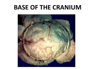
The base of the cranial vault is divided into three cranial fossa: Anterior, middle and posterior Anterior Posterior Middle
Bones of the facial skeleton Zygomatic Maxilla Mandible Nasal Lacrimal Vomer Palatine (hidden)
The sphenoid: seen from behind : its upper part forms the lateral posterior part of orbit: its lower :pterygoid processes support back of maxilla
The nasal bone is a small bone on the midline supporting the top of the nose
Bones of the orbit: These are images of the left orbit: as you look at it the medial side is to the left In upper diagram 1.Frontal bone 2. Ethmoid bone 3. Palatine bone 4.Zygomatic bone 5. Lacrmal bone 6. Maxillary bone The palatine is a small bone extending from the hard palate to the back of the orbit
Upper part of palatine: posterior orbit Lower part of Palatine is posterior part of hard palate Left palatine seen from behind
Orbital fractures • Indirect orbital floor fracture ("blowout fracture") This occurs when the bony rim of the eye remains intact, but the paper thin floor of the eye socket cracks or ruptures. This can cause a small hole in the floor of the eye socket that can trap parts of the eye muscles and surrounding structures. The injured eye may not move normally in its socket, which can cause double vision. Most blowout fractures are caused by an impact to the front of the eye from something bigger than the eye opening, such as a baseball, a fist or an automobile dashboard. F OC SOF N L E Sp Z
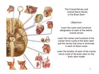
Skull foraminae & fossae Cribiform plate I II Optic canal III, IV, V1,VI SOF V2 f. rotundum f. ovale V3 IAM VII-VIII FM Jugular foramen IX-XI XII Hypoglossal canal
Para-nasal sinuses Frontal Ethmoid Maxillary Sphenoid
Para-nasal air sinuses • 4 pairs that contain mucous membrane & air • Communicate with nasal cavity thro’ foraminae • Act as resonance chambers • Sinusitis infections • thickening of mucosal lining • block entrance to nose • Fluid build-up • Sinus headache • Pain (worse bending forward) F Eth Mx
Para-nasal air sinuses Mx Eth Eth Sph
Lateral wall of the nose Sinus drainage
Normal radiology Middle meningeal artery Lateral view • Used to examine calvarium (vault of skull) • Useful for skull base • You must be able to recognise normal suture lines Coronal suture * PF F E S T M Lamdoid suture Mastoid air cells
Normal radiology AP view • Used for orbit, nasal and paranasal regions CG F SOF Sp E Z T v Mx Mx MD