
The Heart
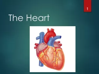
The Heart. The heart=a muscular double pump with 2 functions. Overview. The right side receives oxygen-poor blood from the body and tissues and then pumps it to the lungs to pick up oxygen and dispel carbon dioxide

The Heart
E N D
Presentation Transcript
The heart=a muscular double pump with 2 functions Overview • The right side receives oxygen-poor blood from the body and tissues and then pumps it to the lungs to pick up oxygen and dispel carbon dioxide • Its left side receives oxygenated blood returning from the lungs and pumps this blood throughout the body to supply oxygen and nutrients to the body tissues
simplified… • Cone shaped muscle • Four chambers • Two atria, two ventricles • Double pump – the ventricles • Two circulations • Systemic circuit: blood vessels that transport blood to and from all the body tissues • Pulmonary circuit: blood vessels that carry blood to and from the lungs
Heart’s position in thorax • In mediastinum – behind sternum and pointing left, lying on the diaphragm • It weighs 250-350 gm (about 1 pound) Feel your heart beat at apex (this is of a person lying down)
CXR(chest x ray) Normal male
Chest x rays Lateral (male) Normal female
Starting from the outside… Pericardium(see next slide) Without most of pericardial layers
Layers of the heart wall • Muscle of the heart with inner and outer membrane coverings • Muscle of heart = “myocardium” • The layers from out to in: • Epicardium = visceral layer of serous pericardium • Myocardium = the muscle • Endocardium lining the chambers
Chambers of the heartsides are labeled in reference to the patient facing you • Two atria • Right atrium • Left atrium • Two ventricles • Right ventricle • Left ventricle --------------------------------------------------------------------------------
Chambers of the heartdivided by septae: • Two atria-divided by interatrial septum • Right atrium • Left atrium • Two ventricles-divided by interventricular septum • Right ventricle • Left ventricle
Valvesthree tricuspidone bicuspid (cusp means flap) • “Tricuspid” valve • RA to RV • Pulmonary or pulmonic valve • RV to pulmonary trunk (branches R and L) • Mitral valve (the bicuspid one) • LA to LV • Aortic valve • LV to aorta
Function of semilunar valves (Aortic and pulmonic valves)
Pattern of flow(simple to more detailed) • Body • RA • RV • Lungs • LA • LV • Boby Body to right heart to lungs to left heart to body Body, then via vena cavas and coronary sinus to RA, to RV, then to lungs via pulmonary arteries, then to LA via pulmonary veins, to LV, then to body via aorta From body via SVC, IVC & coronary sinus to RA; then to RV through tricuspid valve; to lungs through pulmonic valve and via pulmonary arteries; to LA via pulmonary veins; to LV through mitral valve; to body via aortic valve then aorta LEARN THIS
Relative thickness of muscular walls LV thicker than RV because it forces blood out against more resistance; the systemic circulation is much longer than the pulmonary circulation Atria are thin because ventricular filling is done by gravity, requiring little atrial effort
Heartbeat Definition: a single sequence of atrial contraction followed by ventricular contraction See http://www.geocities.com/Athens/Forum/6100/1heart.html • Systole: contraction • Diastole: filling • Normal rate: 60-100 • Slow: bradycardia • Fast: tachycardia ***Note: blood goes to RA, then RV, then lungs, then LA, then LV, then body; but the fact that a given drop of blood passes through the heart chambers sequentially does not mean that the four chambers contract in that order; the 2 atria always contract together, followed by the simultaneous contraction of the 2 ventricles
Heart sounds • Called S1 and S2 • S1 is the closing of AV (Mitral and Tricuspid) valves at the start of ventricular systole • S2 is the closing of the semilunar (Aortic and Pulmonic) valves at the end of ventricular systole • Separation easy to hear on inspiration therefore S2 referred to as A2 and P2 • Murmurs: the sound of flow • Can be normal • Can be abnormal
Places to auscultate • Routine places are at right and left sternal border and at apex To hear the sounds: http://www.med.ucla.edu/wilkes/intro.html Note that right border of heart is formed by the RA; most of the anterior surface by the RV; the LA makes up the posterior surface or base; the LV forms the apex and dominates the inferior surface
Cardiac muscle(microscopic) Automaticity: inherent rhythmicity of the muscle itself
“EKG”(or ECG, electrocardiogram) • Electrical depolarization is recorded on the body surface by up to 12 leads • Pattern analyzed in each lead P wave=atrial depolarization QRS=ventricular depolarization T wave=ventricular repolarization
specialized cardiac muscle cells that carry impulses throughout the heart musculature, signaling the chambers to contract in the proper sequence Electrical conduction system: (Explanation in next slides)
Conduction system • SA node (sinoatrial) • In wall of RA • Sets basic rate: 70-80 • Is the normal pacemaker • Impulse from SA to atria • Impulse also to AV node via internodal pathway • AV node • In interatrial septum
Conduction continued • SA node through AV bundle (bundle of His) • Into interventricular septum • Divides R and L bundle branches become subendocardial branches (“Purkinje fibers”) • Contraction begins at apex
Autonomic innervation • Sympathetic • Increases rate and force of contractions • Parasympathetic (branches of Vagus n.) • Slows the heart rate For a show on depolarization: http://education.med.nyu.edu/courses/old/physiology/courseware/ekg_pt1/EKGseq.html
Blood supply to the heart(there’s a lot of variation) A: Right Coronary Artery; B: Left Main Coronary Artery; C: Left Anterior Descending (LAD, or Left Anterior Interventricular);D: Left Circumflex Coronary Artery; G: Marginal Artery; H: Great Cardiac Vein; I: Coronary sinus, Anterior Cardiac Veins.
A lot of stuff from anterior view Each atrium has an “auricle,” an ear-like flap
Again posterior view Note: the coronary sinus (largest cardiac vein) – delivers blood from heart wall to RA, along with SVC & IVC)


