Exploring Microscopy Techniques for Cell Observation
900 likes | 929 Vues
Discover the principles of light and electron microscopy, techniques such as staining, and the morphology of prokaryotic cells in this comprehensive chapter on cell structure observation methods.

Exploring Microscopy Techniques for Cell Observation
E N D
Presentation Transcript
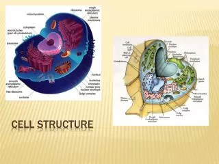
Microscopy and Cell Structure Chapter 3 Part I Observing Cells
Principles of Light Microscopy • Light Microscopy • Most common and easiest to use: bright-field microscope • Important factors in light microscopy include • Magnification • Resolution • Contrast
Principles of Light Microscopy • Magnification • two magnifying lenses • Ocular lens and objective lens • condenser lens • focus illumination on specimen
Principles of Light Microscopy • Resolution • minimum distance between two objects that still appear as separate objects • determine the usefulness of microscope
Principles of Light Microscopy • Factors affect resolution • Lens • Wavelength of light • How much light is released from the lens • magnification • Maximum resolving power of most brightfield microscopes is 0.2 μm (1x10⁻⁶) • sufficient to see most bacteria • Too low to see viruses
Principles of Light Microscopy • Resolution is enhanced with lenses of higher magnification (100x) by the use of immersion oil • Oil reduces light refraction • Immersion oil has nearly same refractive index as glass
Principles of Light Microscopy • Contrast • Reflects the number of visible shades in a specimen • increase contrast • Use special microscopes • specimen staining
Principles of Light Microscopy • Examples of light microscopes that increase contrast • Phase-Contrast Microscope • Interference Microscope • Dark-Field Microscope • Fluorescence Microscope • Confocal Scanning Laser Microscope
Principles of Light Microscopy • Phase-Contrast • Amplifies differences between refractive indexes of cells and surrounding medium • Darker appearance for denser materials. • Uses set of rings and diaphragms to achieve resolution
Principles of Light Microscopy • Interference Scope • appear three dimensional • Depends on differences in refractive index
Principles of Light Microscopy • Dark-Field Microscope • Reverse image • Like a photographic negative • a modified condenser directs the lights at an angle and only the light scattered by the specimen enters the objective lens
Principles of Light Microscopy • Fluorescence Microscope • observe organisms naturally fluorescent or flagged with fluorescent dye • Fluorescent molecule absorbs ultraviolet light and emits visible light • Image fluoresces on dark background
Principles of Light Microscopy • Electron Microscope • Uses electromagnetic lenses, electrons and fluorescent screen to produce image • Resolution increased 1,000 fold over brightfield microscope • To about 0.3 nm (1x10⁻⁹) • Magnification increased to 100,000x • Two types of electron microscopes • Transmission • Scanning
Quiz • With 10x ocular lens and 40x objective lens, what is the magnifying power?
Quiz • What are the three important factors for microscope?
Microscope TechniquesDyes and Staining • Dyes and Staining • stained to observe organisms • made of organic salts • Basic dyes carry positive charge • Acidic dyes carry negative charge
Microscope TechniquesDyes and Staining • Common basic dyes include • Methylene blue • Crystal violet • Safrinin • Malachite green
Microscope TechniquesDyes and Staining • Simple staining • use one color to stain • increase contrast between cell and background
Microscope TechniquesDyes and Staining • Differential Stains • to distinguish one bacterial group from another • Uses a series of reagents • Two most common differential stains • Gram stain • Acid-fast stain
Microscope TechniquesDyes and Staining • Gram Stain • widely used procedure for classiffying bacteria • two major groups based on cell wall structural differences • Gram positive • Gram negative
Microscope TechniquesDyes and Staining • Gram Stain • Involves four reagents • Primary stain • Mordent • Decolorizer • Counter or Secondary stain Old gram positive appears to be gram negative
Microscope TechniquesDyes and Staining • Acid-fast Stain • Used to stain members of genus Mycobacterium • High lipid concentration in cell wall • Uses heat to facilitate staining
Microscope TechniquesDyes and Staining • Acid-fast Stain • used for presumptive identification in diagnosis of clinical specimens • Requires multiple steps • Primary dye • Decolorizer • Counter stain
Microscope TechniquesDyes and Staining • Special Stains • Capsule stain • Endospore stain • Uses heat to facilitate staining • Flagella stain
Quiz • What are the two commonly used differential staining method?
Morphology of Prokaryotic Cells • Prokaryotes exhibit a variety of shapes • Coccus • Bacillus • Do not to be confused with Bacillus genus
Morphology of Prokaryotic Cells • Coccobacillus • Vibrio • Spirillum • Spirochete • Pleomorphic
Morphology of Prokaryotic Cells • groupings morphology • Cells adhere together after cell division for characteristic arrangements • Especially in the cocci
Morphology of Prokaryotic Cells • Division along a single plane may result in pairs or chains of cells • Pairs = diplococci • Example: Neisseria gonorrhoeae • Chains = streptococci • Example: species of Streptococcus
Morphology of Prokaryotic Cells • Division along two or three perpendicular planes form cubical packets • Example: Sarcina genus • Division along several random planes form clusters • Example: species of Staphylococcus
Microscope • Three important factors • Staining. • Prokaryotic morphology
Microscopy and Cell Structure Part II - Prokaryotic Cell Structure
Cytoplasmic membrane • Defines the boundary of the cell • Semi-permeable; • Transport proteins function as selective gates (selectively permeable) • Control entrance/expulsion of antimicrobial drugs • Receptors provide a sensor system • Phospholipid bilayer, embedded with proteins
Cytoplasmic membrane • Defines the boundary of the cell • Semi-permeable; • Transport proteins function as selective gates (selectively permeable) • Control entrance/expulsion of antimicrobial drugs • Receptors provide a sensor system • Phospholipid bilayer, embedded with proteins
Cytoplasmic membrane • Defines the boundary of the cell • Semi-permeable; • Transport proteins function as selective gates (selectively permeable) • Control entrance/expulsion of antimicrobial drugs • Receptors provide a sensor system • Phospholipid bilayer, embedded with proteins • Fluid mosaic model
Cytoplasmic Membrane • Methods for molecule to go cross membrane • Simple diffusion: the only system does not rely on transport protein • Facilitated diffusion • Active transport • Group transport
Cytoplasmic Membrane • Simple diffusion- • Water, certain gases and small hydrophobic molecules • Move along with concentration gradient • Osmosis
Cytoplasmic Membrane • Movement of molecules across membrane by transport systems • Specific Transport systems include • Facilitated diffusion • Active transport • Group translocation
Directed Movement of Molecules Across the Cytoplasmic Membrane Facilitated diffusion no energy expended
Directed Movement of Molecules Across the Cytoplasmic Membrane Facilitated diffusion Active transport - energy is expended • Moves compounds against a concentration gradient
Major facilitator superfamily (expends proton motive force) ABC transport systems (expends ATP) Directed Movement of Molecules Across the Cytoplasmic Membrane Facilitated diffusion Active transport - energy is expended • Use binding proteins to scavenge and deliver molecules to transport complex • Example: maltose transport • Example: efflux pumps used in antimicrobial resistance
Cytoplasmic membrane Proton: H+ Proton motive force: Energy stored in the electrochemical gradient created by electron transport chain Electron transport chain Electron transport chain - Series of proteins that sequentially transfer electrons and eject protons from the cell, creating an electrochemical gradient • Proton motive force is used to fuel: • Synthesis of ATP (the cell’s energy currency) • Rotation of flagella (motility) • One form of transport
Directed Movement of Molecules Across the Cytoplasmic Membrane Facilitated diffusion Active transport - Chemically modifies a compound during transport Group translocation
Directed Movement of Molecules Across the Cytoplasmic Membrane Facilitated diffusion Active transport Group translocation Secretion - Transport of proteins to the outside • Characteristic sequence of amino acids in a newly synthesized protein functions as a tag (signal sequence)
Prokaryotic structure • Cell membrane structure • Movements across membrane
Cell Wall Provides rigidity to the cell (prevents it from bursting)
Cell Wall • Bacterial cell wall • Rigid structure • Determines shape of bacteria • Protection • Unique chemical structure • Distinguishes Gram positive from Gram-negative
Cell Wall • Peptidoglycan- rigid molecule; unique to bacteria • Alternating subunits of NAG and NAM form glycan chains • Glycan chains are connected to each other via peptide chains on NAM molecules