Neural Processing and Communication: Understanding Brain Structures and Functions
Explore the intricate neural structures of the brain, including the motor cortex, visual cortex, and sensory associative cortex. Understand the mechanisms of neural computation, thought, and communication. Discover how neurons communicate and process information.

Neural Processing and Communication: Understanding Brain Structures and Functions
E N D
Presentation Transcript
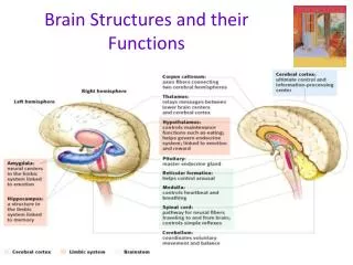
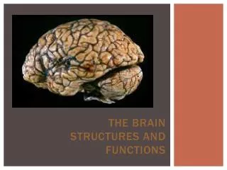
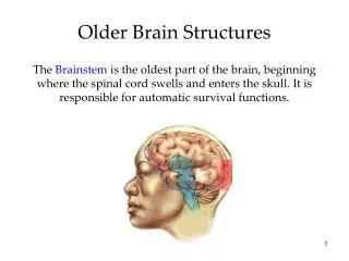
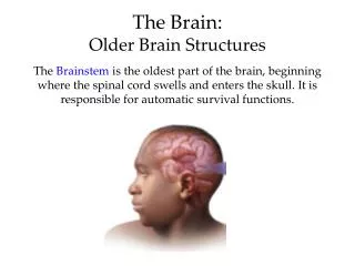
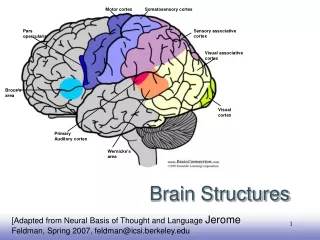
Motor cortex Somatosensory cortex Sensory associative cortex Pars opercularis Visual associative cortex Broca’s area Visual cortex Primary Auditory cortex Wernicke’s area Brain Structures [Adapted from Neural Basis of Thought and Language Jerome Feldman, Spring 2007, feldman@icsi.berkeley.edu
Intelligence Learning and Understanding • I hear and I forget • I see and I remember • I do and I understand • attributed to Confucius 551-479 B.C. • There is no erasing in the brain
Intelligence and Neural Computation • What it means for the brain to compute and how that computation differs from the operation of a standard digital computer. • How intelligence can be implemented in the structure of the neural circuitry of the brain. • How is thought related to perception, motor control, and our other neural systems, including social cognition? • How do the computational properties of neural systems and the specific neural structures of the human brain shape the nature of thought? • What are the applications of neural computing?
Nervous System Divisions • Central nervous system (CNS) • brain • spinal cord
Nervous System Divisions • Peripheral nervous system (PNS) consists of: • Cranial and spinal nerves • Ganglia • Sensory receptors • Subdivided into: • Somatic • Autonomic • Motor component subdivided into: • sympathetic • parasympathetic • Enteric
1000 operations/sec 100,000,000,000 units 10,000 connections/ graded, stochastic embodied fault tolerant evolves learns 1,000,000,000 ops/sec 1-100 processors ~ 4 connections binary, deterministic abstract crashes designed programmed Brains ~ Computers
Neurons • cell body • dendrites (input structure) • receive inputs from other neurons • perform spatio-temporal integration of inputs • relay them to the cell body • axon (output structure) • a fiber that carries messages (spikes) from the cell to dendrites of other neurons
Neuron cells • unipolar • bipolar • multipolar
science-education.nih.gov Synapse • site of communication between two cells • formed when an axon of a presynaptic cell “connects” with the dendrites of a postsynaptic cell
Synapse axon of presynaptic neuron dendrite of postsynaptic neuron bipolar.about.com/library
Synapse • a synapse can be excitatory or inhibitory • arrival of activity at an excitatory synapsedepolarizesthe local membrane potential of the postsynaptic cell and makes the cell more prone to firing • arrival of activity at an inhibitory synapsehyperpolarizes the local membrane potential of the postsynaptic cell and makes it less prone to firing • the greater the synaptic strength, the greater the depolarization or hyperpolarization
Visual cortex of the rat
Somatotopy of Action Observation Foot Action Hand Action Mouth Action Buccino et al. Eur J Neurosci 2001
Amoeba eating Artist’s rendition of a typical cell membrane
Neural Processing From lecture notes by Dr Rachel Swainson NEURAL COMMUNICATION 1: Transmission within a cell and from a lecture notes based on www.unisanet.unisa.edu.au/Information/12924info/Lecture Presentation - Nervous tissue.ppt
Transmission of information Information must be transmitted • within each neuron • and between neurons
The Membrane • The membrane surrounds the neuron. • It is composed of lipid and protein.
Cell Electrical Potential Every neuron is covered by a membrane The membrane is selectively permeable to the passage of chemical molecules (ions) The membrane maintains a separation of electrical charge across the cell membrane. The cell membrane has an electrical potential Electrical potentials Electrical charge of the membrane is related to charged ion that cross the membrane through lipids, ion channels and protein ion-transporters. Electrical currents (ionic flux) The flow of electrical charge between the cell’s interior and exterior cellular fluids
Forces determine flux of ions • Electrostatic forces • Particles with opposite charges attract, Identical charges repel • Concentration forces • Diffusion – molecules distribute themselves evenly – • Protein – ion channels • Selective Non – gated ion channels • Selective Voltage-dependent gated ion channels • Protein – ion transporters • K+ Na + pump • Cl - pump
outside + + + + + - inside - - - - Resting potential of neuron = -70mV The Resting Potential • There is an electrical charge across the membrane. • This is the membrane potential. • The resting potential (when the cell is not firing) is a 70mV difference between the inside and the outside.
Ions and the Resting Potential • Ions are electrically-charged molecules e.g. sodium (Na+), potassium (K+), chloride (Cl-). • The resting potential exists because ions are concentrated on different sides of the membrane. • Na+ and Cl- outside the cell. • K+ and organic anions inside the cell. Cl- Na+ Cl- Na+ Na+ Na+ outside inside Organic anions (-) K+ Organic anions (-) Organic anions (-) K+
Maintaining the Resting Potential • Na+ ions are actively transported (this uses energy) to maintain the resting potential. • The sodium-potassium pump (a membrane protein) exchanges three Na+ ions for two K+ ions. Na+ Na+ Na+ outside inside K+ K+
Neuronal firing: the action potential • The action potential is a rapid depolarization of the membrane. • It starts at the axon hillock and passes quickly along the axon. • The membrane is quickly repolarized to allow subsequent firing.
Course of the Action Potential • The action potential begins with a partial depolarization (e.g. from firing of another neuron ) [A]. • When the excitation threshold is reached there is a sudden large depolarization [B]. • This is followed rapidly by repolarization [C] and a brief hyperpolarization [D].
The Action Potential • The action potential is “all-or-none”. • It is always the same size. • Either it is not triggered at all - e.g. too little depolarization, or the membrane is “refractory”; • Or it is triggered completely.
Action potential 2 phases: • Depolarisation • graded potentials move toward firing threshold • if reach threshold voltage regulated sodium channels open • reversal of membrane permeability • Repolarisation • sodium channels close • potassium channels open
Na+ + - - + Na+ Na+ Action potentials: Rapid depolarization • When partial depolarization reaches the activation threshold,voltage-gated sodium ion channels open. • Sodium ions rush in. • The membrane potential changes from -70mV to +40mV.
Na+ K+ + K+ - Na+ Na+ K+ Action potentials: Repolarization • Sodium ion channels close and become refractory. • Depolarization triggers opening of voltage-gated potassium ion channels. • K+ ions rush out of the cell, repolarizing and then hyperpolarizing the membrane.
Conduction of the action potential • Passive conduction will ensure that adjacent membrane depolarizes, so the action potential “travels” down the axon. • But transmission by continuous action potentials is relatively slow and energy-consuming (Na+/K+ pump). • A faster, more efficient mechanism has evolved: saltatory conduction. • Myelination provides saltatory conduction.
Propagation of the Action Potential • Action Potential spreads down the axon in a chain reaction • Unidirectional • it does not spread into the cell body and dendrite due to absence of voltage-gated channels there • Refraction prevents spread back across axon
Myelination • Most mammalian axons are myelinated. • The myelin sheath is provided by oligodendrocytes and Schwann cells. • Myelin is insulating, preventing passage of ions over the membrane.
Saltatory Conduction • Myelinated regions of axon are electrically insulated. • Electrical charge moves along the axon rather than across the membrane. • Action potentials occur only at unmyelinated regions: nodes of Ranvier. Myelin sheath Node of Ranvier
Summary of axonal conduction • Unmyelinated fibres • continuous conduction • Myelinated fibres • saltatory conduction • High density of voltage gated channels at Nodes of Ranvier • Larger diameter axons propagate impulses faster • Stimulus intensity encoded by: • frequency of impulse generation • number of sensory neurons activated
Synaptic transmission • Information is transmitted from the presynaptic neuron to the postsynaptic cell. • Chemical neurotransmitters cross the synapse, from the terminal to the dendrite or soma. • The synapse is very narrow, so transmission is fast.
terminal extracellular fluid synaptic cleft presynaptic membrane dendritic spine postsynaptic membrane Structure of a synapse • An action potential causes neurotransmitter release from the presynaptic membrane. • Neurotransmitters diffuse across the synaptic cleft. • They bind to receptors within the postsynaptic membrane, altering the membrane potential.
Neurotransmitter release • Synaptic vesicles, containing neurotransmitter, congregate at the presynaptic membrane. • The action potential causes voltage-gated calcium (Ca2+) channels to open; Ca2+ ions flood in. vesicles Ca2+ Ca2+ Ca2+ Ca2+ Ca2+ Ca2+ Ca2+ Ca2+
Ca2+ Neurotransmitter release • Ca2+ causes vesicle membrane to fuse with presynaptic membrane. • Vesicle contents empty into cleft: exocytosis. • Neurotransmitter diffuses across synaptic cleft.
ACh ACh Ionotropic receptors • Synaptic activity at ionotropic receptors is fast and brief (milliseconds). • Acetyl choline (Ach) works in this way at nicotinic receptors. • Neurotransmitter binding changes the receptor’s shape to open an ion channel directly.
Ionotropic Receptors 4 nm