Download
1 / 1
10 likes | 117 Vues
This study presents evidence of a previously unrecognized strong left-right asymmetric segmentation bias in subcortical brain structures. This bias significantly impacts left-right asymmetry analyses in neuroimaging, indicating that many existing studies need reconsideration. Our findings highlight variable segmentation methods and suggest that care must be taken in interpreting hemispheric asymmetry, especially with structures like the caudate, putamen, and amygdala. The work emphasizes the necessity for improved segmentation protocols to mitigate such biases in future analyses.

E N D
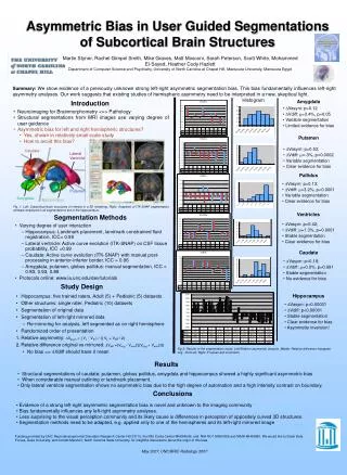
Asymmetric Bias in User Guided Segmentations of Subcortical Brain Structures Martin Styner, Rachel Gimpel Smith, Mike Graves, Matt Mosconi, Sarah Peterson, Scott White, Mohammed El-Sayed, Heather Cody Hazlett Department of Computer Science and Psychiatry, University of North Carolina at Chapel Hill, Mansoura University, Mansoura Egypt NIAL Summary: We show evidence of a previously unknown strong left-right asymmetric segmentation bias. This bias fundamentally influences left-right asymmetry analyses. Our work suggests that existing studies of hemispheric asymmetry need to be interpreted in a new, skeptical light. Histogram Amygdala Introduction • Vasym: p=0.12 • Vdiff: =3.4%, p=0.05 • Variable segmentation • Limited evidence for bias • Neuroimaging for Brainmorphometry <=> Pathology • Structural segmentations from MRI images use varying degree of user guidance • Asymmetric bias for left and right hemispheric structures? • Yes, shown in relatively small scale study • How to avoid this bias? Putamen • Vasym: p=0.53; • Vdiff: =-3%, p=0.0002 • Variable segmentation • Clear evidence for bias Caudate Lateral Ventricle Pallidus • Vasym: p=0.13; • Vdiff: =3.2%, p<0.0001 • Variable segmentation • Clear evidence for bias GP Putamen Amygdala Hippocampus Fig. 1. Left: Subcortical brain structures of interest in a 3D rendering. Right: Snapshot of ITK-SNAP segmentation software employed in all segmentations but of the hippocampus. Ventricles Segmentation Methods • Vasym: p=0.42; • Vdiff: =1.3%, p=0.0001 • Stable segmentation • Clear evidence for bias • Varying degree of user interaction • Hippocampus: Landmark placement, landmark constrained fluid registration, ICC= 0.99 • Lateral ventricle: Active curve evolution (ITK-SNAP) on CSF tissue probability, ICC =0.99 • Caudate: Active curve evolution (ITK-SNAP) with manual post-processing in anterior-inferior border, ICC = 0.96 • Amygdala, putamen, globus pallidus: manual segmentation, ICC = 0.83, 0.93, 0.89 • Protocols online: www.ia.unc.edu/dev/tutorials Caudate • Vasym: p=0.18; • Vdiff: =0.0%, p=0.991 • Stable segmentation • No evidence for bias Study Design Hippocampus • Hippocampus: five trained raters, Adult (5) + Pediatric (5) datasets • Other structures: single rater, Pediatric (10) datasets • Segmentation of original data • Segmentation of left-right mirrored data • Re-mirroring for analysis, left segmented as on right hemisphere • Randomized order of presentation • Relative asymmetry: Vasym = ( VL - VR ) / (( VL + VR) / 2) • Relative difference original vs mirrored: Vdiff =(Vorig - Vmirr)/((Vorig + Vmirr)/2) • No bias => Vdiff should have 0 mean • Vasym: p<0.00001 • Vdiff: p<0.00001 • Stable segmentation • Clear evidence for bias • Asymmetry inversion! Fig 2. Results of the segmentation study: Left:Relative asymmetry analysis, Middle: Relative difference histogram orig - mirrored, Right: P-values and comments. Results • Structural segmentations of caudate, putamen, globus pallidus, amygdala and hippocampus showed a highly significant asymmetric bias • When considerable manual outlining or landmark placement. • Only lateral ventricle segmentation shows no asymmetric bias due to the high degree of automation and a high intensity contrast on boundary. Conclusions • Evidence of a strong left-right asymmetric segmentation bias is novel and unknown to the imaging community • Bias fundamentally influences any left-right asymmetry analyses. • Less surprising to the visual perception community and its likely cause is differences in perception of oppositely curved 3D structures. • Segmentation methods need to be adapted, e.g. applied only to one of the hemispheres and its left-right mirrored image Funding provided by UNC Neurodevelopmental Disorders Research Center HD 03110, the NIH Conte Center MH064065, and NIH RO1 MH61696 and NIMH MH64580. We would like to thank Dale Purves, Duke University, and Donald Mershon, North Carolina State University, for insightful discussions about the origin of this bias. May 2007, UNC/BRIC Radiology 2007