Download
1 / 10
100 likes | 130 Vues
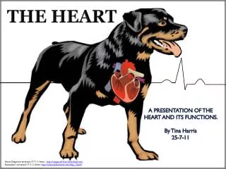
Learn about the heart components like pericardium, heart wall, chambers, valves, and essential blood vessels. Explore blood flow patterns with informative videos.

E N D
Membranes • Pericardium • Covers the heart • Visceral – innermost layer • Parietal – outermost layer • Pericardial fluid: serous fluid found between visceral & parietal layers reduces friction
Heart Wall • Epicardium (visceral pericardium): outer covering • Myocardium: cardiac muscle tissue • Endocardium: epithelial & connective tissue; continuous with inner linings of blood vessels attached to heart
A: Superior Vena Cava B: R. Pulmonary Artery C: R. Pulmonary Vein D: R. Atrium E: Tricuspid Valve F: R. Ventricle G: Inferior Vena Cava H: Myocardium (Septum) I: L. Ventricle J: Bicuspid Valve K: Pulmonary Valve L: L. Atrium M: L. Pulmonary Vein N: Pulmonary Trunk O: Aorta
Chambers • Atria (right & left): thin walls, upper chambers; receive blood returning to the heart • Ventricles (right & left): lower chambers; receive blood from atria, contract to force blood into arteries • Septum: divides heart into right & left sides
Valves • 2 Artioventricular (AV) valves: prevent backflow of blood into atria • Tricuspid valve: between RA & RV • Bicuspid Valve: between LA & LV • Chordae tendineae = fibrous strings that attach from papillary muscles to valve; open valves • Papillary muscles = small mounds of cardiac muscle tissue • Aortic valve: prevents backflow of blood into L ventricle from aorta • Pulmonary Valve: prevents backflow of blood into R ventricle from pulmonary trunk
Essential Blood Vessels • Pulmonary trunk: divides to form R/L pulmonary arteries, which carry blood into lungs from R ventricle • R/L pulmonary veins: carry blood from lungs to L atrium • Aorta: large artery, carries blood away from heart to body
Khan Academy Video: • https://www.youtube.com/watch?v=7XaftdE_h60 • Fetal Heart https://www.youtube.com/watch?v=cgccQVcFLi4