Fluoroscopy Safety
Fluoroscopy Safety . New Wisconsin Regulations. In 2010, WI enacted new training regulations for clinicians who use fluoroscopy. Unless certified by the American Board of Radiology (or board eligible), clinicians are required to be trained in:
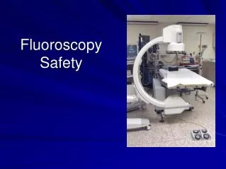

Fluoroscopy Safety
E N D
Presentation Transcript
New Wisconsin Regulations In 2010, WI enacted new training regulations for clinicians who use fluoroscopy. Unless certified by the American Board of Radiology (or board eligible), clinicians are required to be trained in: Principles of operation of the fluoroscopic x-ray system Biological effects of x-rays Principles of radiation protection Fluoroscopic outputs High Level control options Dose reduction techniques Applicable state and federal regulations
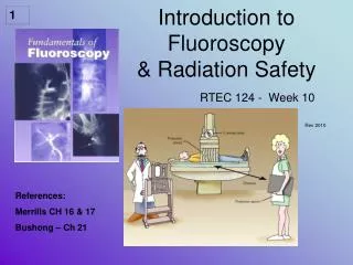
What is Fluoroscopy? Fluoroscopy is an imaging procedure that uses a continuous x-ray beam to create real-time images viewed on a monitor. It enables physicians to view internal organs and vessels in motion. Fluoroscopy is used in both diagnostic and therapeutic procedures.
Medical uses of fluoroscopybegan shortly after Roentgen’sdiscovery of x-rays in 1895. Fluoroscopy Today Fluoroscopy for tuberculosis (1940)
1990’s: Injuries Reported to FDA From 1992 through 1995, the FDA received more than 100 reports of patients with radiation injuries from fluoroscopy. Since 1992, reports of injuries to patients and physicians have appeared in radiology, cardiology, and medical physics journals.
Skin Injury and Time to OnsetListed in order of time of initial onset
Skin Injury and Time to Onset Source: Centers for Disease Control and Prevention. Cutaneous radiation injury: fact sheet for physicians.
Example 1 A 40-year-old male underwent coronary angiography, coronary angioplasty and a second angiography procedure due to complications, followed by a coronary artery by-pass graft, all on March 29, 1990. Example and images provided by Thomas Shope, U.S. FDA Center for Devices and Radiological Health
6-8 weeks post procedure Note the erythema in the shape of the radiation collimation
16-21 weeks post procedure Erythema reduced, Secondary Damage (not as well imaged)
18-21 months post procedure Close-up view of lesion
Note Epilation Example 2 Injury following three procedures involving transjugularintrahepaticportosystemic shunt placement (TIPS), demonstrating disfigurement after surgical correction. Koenig TR, Wolff D, Mettler FA et al. Skin injuries from fluoroscopically guided procedures: part 1, characteristics of radiation injury. AJR Am J Roentgenol 2001; 177(1):3-11.
Example 3 Injury to arm of patient. Patient was draped for procedure and physicians did not realize that she had moved her arm so that it was resting on the port of the X-ray tube during the procedure. Wagner LK, Archer BR. Minimizing Risks from Fluoroscopic X Rays. 4th edition. The Woodlands, Texas: Partners in Radiation Management, 2004.
Why Are Injuries Occurring? One contributing factor is the growth in number and types of interventional procedures using fluoroscopy. But any procedure using fluoroscopy has the potential for patient injury. Another factor may be more overweight and obese patients. Higher energy x-rays and higher radiation dose rates are required to penetrate through these patients.
FDA Actions In 1994, the FDA issued a Public Health Advisory on avoidance of serious skin injuries to patients during fluoroscopy-guided procedures. In 1995, the FDA issued a follow-up advisory on recording information in the patient’s record that identifies the potential for serious skin injury from fluoroscopy.
Joint Commission Action In 2006, the Joint Commission added a Sentinel Event category for radiation overdose involving prolonged fluoroscopy with a cumulative dose of more than 15 Gray to a single field. Fluoroscopy machines manufactured after June 2006 measure and display a reference patient radiation dose. The reference dose can be monitored during the procedure, and the cumulative dose can be recorded in the patient’s medical record.
Summary All of the following injuries can be caused by radiation: • Skin erythema and desquamation • Epilation • Skin ulceration
What About Personnel Safety? Physicians and staff using fluoroscopy are exposed to: - Scattered radiation from the patient • Leakage radiation from the x-ray tube • Primary radiation from the x-ray beam if their hands are in the radiation field Detector/image intensifier x-ray tube
Personnel Safety Although clinician radiation dose is much lower than the patient dose, it is proportional to patient dose. Higher patient doses will usually lead to higher operator and staff doses.
Radiation Risks High doses of radiation (>1 Gray in a single exposure), such as those received by patients injured by fluoroscopy, are linked to skin injury and increased risk of cancer. Low doses of radiation over long periods of time, such as those received by medical personnel, may result in an increased risk of cancer, although this has not been conclusively proven.
ALARA (As Low As Reasonably Achievable) Because we know that large doses of radiation can cause long term health effects, such as increasing the risk of developing cancer, we assume that all radiation exposure entails some risk. Therefore, we should try to limit the radiation exposure to patients and staff, consistent with obtaining the necessary clinical information.
In fluoroscopy, there are three practical techniques to reduce radiation exposure to patients and personnel. • Reduce Fluoro Time • Increase Distance • Provide Shielding The following slides demonstrate how to use these techniques to reduce radiation exposure.
Time: Identify if the patient has had other recent long fluoro procedures Check the patient’s medical record to see if they have had a recent long fluoroscopy procedure in the same location. If yes, try to change the C-Arm angle so that you are not irradiating the same area of skin again.
Time: Recognize the Fluoroscopy “Beam-On” Controls Typical x-ray “beam-on” foot pedal. Most units also have a beam-on button or switch the user can operate by hand.
Time: Minimize “Beam-On” time Use short taps of the fluoroscopy beam-on control. Don’t use a “lead foot” on the fluoroscopy pedal. Reducing beam-on time is the most effective way to reduce dose.
Time: LIH and LFH Use Last Image Hold (LIH) or Last Fluoroscopy Hold (LFH) when possible instead of re- exposing the patient. Last Image Hold saves the last fluoroscopy image and displays it on the monitor. Last Fluoroscopy Hold saves the last video sequence of fluoroscopy images for instant replay.
Time: Fluoroscopy Dose Modes Different dose mode selections may be available • Low Dose (↓patient dose, ↑ image noise) • High Dose (↑patient dose, ↓ image noise) • Low Frame Rate (↓patient dose,↓ frame rate) When Image Quality allows, use low dose mode and/or a lower frame rate.
Time: Minimize Use of High Dose Mode High dose rate mode may be needed for large patients or for seeing greater detail. High dose mode selection is usually denoted by a “+” sign. Do not routinely use high dose mode.
Time: Digital Acquisition Mode X-Ray machines used for interventional procedures have a digital acquisition or “cine” mode. A high radiation dose rate is used to obtain a series of high resolution images with reduced image noise. The radiation dose per frame for digital acquisitions can be 15 times greater than for fluoroscopy.
Time: Use Digital Acquisition/Cine Mode Appropriately The number and length of digital acquisition or cine “runs” may be the greatest source of patient radiation dose in interventional radiology procedures. Be aware of the increased dose rate and do not use digital acquisition/cine mode as a substitute for fluoroscopy.
Using Time to Reduce Exposure: Summary When image quality allows, choosing to use low dose fluoro modes and last image hold, while limiting the use of “boost” fluoro and high dose digital acquisitions, will reduce patient and staff radiation exposure.
Distance: Scattered Radiation During fluoroscopy, radiation is scattered from the surface of the patient where the x-ray beam enters. Scattered radiation is the main source of radiation dose to staff. It also decreases image contrast and degrades image quality. Detector/Image Intensifier x-ray tube
Distance: C-Arm Position Position the X-ray tube underneath the patient, not above the patient. The greatest amount of scatter radiation is produced where the x-ray beam enters the patient. By positioning the x-ray tube below the patient, you receive less scatter radiation. Image Intensifier X-ray Tube
Distance: C-Arm Position Always stand closer to the detector/image intensifier. For lateral and oblique projections, position the C-arm so that the x-ray tube is on the opposite side of the patient from where you are working. This will reduce the scatter radiation reaching you. Always stand farther from the X-Ray Tube.
Distance: C-Arm Position Position the x-ray tube and image intensifier so you are working on the image intensifier side of the patient. Position the x-ray tube as far from the patient as possible. Position the Image intensifier as close to the patient as possible. X-ray tube Image intensifier
Distance: Proximity to the X-Ray Tube The patient’s skin should never touch or be near the x-ray tube port (where the x-rays come out). Staff should also never touch or be near the x-ray tube port. Burns can occur in seconds if skin is touching or near the x-ray tube port. X-ray tube port
Distance: Minimize the Air Gap Move the detector or image intensifier as close to the patient as possible. A smaller air gap reduces radiation dose to the patient and staff and improves image quality.
Distance: When possible increase your distance from the patient when the x-ray beam is on When possible, simply taking a step back from the radiation source whenever possible will greatly reduce your radiation dose. Moving from 30cm to 60 cm from the patient will reduce your exposure by a factor of 4.
Distance: Stay Out of the Fluoroscopy Beam Don’t put your hands in the fluoroscopy beam unless absolutely necessary for the procedure. This is the hand of a physician who was exposed to repeated small doses of x-ray radiation for 15 years. The skin cancer appeared several years after his work with x-rays had ceased. Meissner, William A. and Warren, Shields: Neoplasms, In Anderson W.A.D. editor; Pathology, edition 6, St. Louis, 1971, The C.V. Mosby Co
Using Distance to Reduce Exposure: Summary • When possible, always position the image intensifier under the patient. • Maximize the distance from the x-ray tube to the patient. • Move the image intensifier as close to the patient as you can. • Maximize the distance between you and the patient during the x-ray exposure. • Do not put your hands in the primary beam.
Shielding: Collimate Appropriately Collimate tightly to the area of clinical interest to reduce patient and staff dose, reduce scatter, and improve image contrast. uncollimated collimated
Shielding: Magnification Modes Magnification enlarges the anatomy being viewed, but it also increases the radiation dose to the patient. Multiple electronic magnification modes may be available.
Use Shielding Wisconsin DHS regulations require anyone within 6 feet of a fluoroscopy machine to wear a lead apron. You may also wear a lead thyroid shield or leaded eyeglasses, depending on the type and amount of work you do.
Shielding: Mini C-Arms Although Mini C-Arms produce less scatter Radiation than full C-Arms, Aspirus Wausau Hospital radiation safety procedures require the use of lead aprons when performing any fluoroscopy procedure. GE OEC Mini-C
Shielding: Hang Lead Aprons Properly Hanging lead aprons on hangers/hooks prevents the lead from cracking and tearing. This is for your safety, so please be sure to take care of your lead.
Using Shielding to Reduce Exposure: Summary • Collimate the radiation to the area of interest. • Minimize the use of high magnification modes. • Always wear radiation protection devices.
Pediatric Patients Children are estimated to be two to seven times more sensitive to radiation than adults. They have more dividing and differentiating cells and have a longer time over which radiation effects such as cancer can appear. Use techniques taught in this course to minimize the dose to pediatric patients as well.