Download
1 / 47
470 likes | 587 Vues
HISTOLOGY The Study of Tissues. Tissue Types. Epithelial, Connective, Muscular, Nervous Joined into functioning units by points of contact between cell membranes of adjoining cells ( cell junctions )

E N D
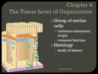
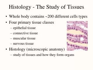
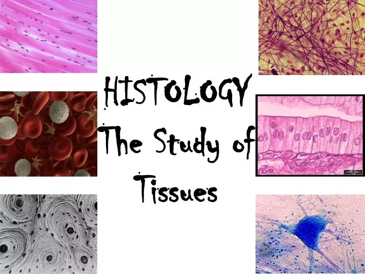
HISTOLOGY The Study of Tissues
Tissue Types • Epithelial, Connective, Muscular, Nervous • Joined into functioning units by points of contact between cell membranes of adjoining cells (cell junctions) • Cell junctions prevent movement between cells, prevent separation during function, allow communication
Epithelial Tissue • Coverings and linings – forms outer coverings of skin and some organs, lines body cavities, blood vessels, ducts, and interiors of reproductive, respiratory, digestive, and urinary organs, makes up parts of sense organs (hearing, vision, and touch) • Glandular – internal and external secretion portions of glands
General Features - Epithelial Tissues • Closely packed cells with little extracellular material between them • Arranged in continuous sheets, single or double layers • Apical (free) surface – exposed to a body cavity or an exterior surface; most superficial • Lateral surface – faces adjacent cells • Basal surface – deepest layer • Basement membrane – extracellular structure composed mostly of fibers. Between epithelial layers and connective tissue. Binds the 2 tissues together for support
General Features – Epithelial Tissue • Avascular (no blood supply) within the tissue. Blood vessels bringing in nutrients/O2 and removing waste/CO2 located in nearby connective tissue. Nutrients and wastes move in and out of epithelial tissue via diffusion (passive hi to lo) • Have a nerve supply • High capacity for reproduction due to wear, tear, and injury
Types of Epithelium – Simple Squamous • Thin, flat cells allows rapid movement of materials through cells • Form “solid” surface • Nucleus is centrally located and flattened oval • Found in areas of body where rapid filtering, diffusion, absorption, and secretion are necessary (kidneys, lungs, linings of heart, blood vessels, lymph vessels) • Not subjected to wear and tear
Types of Epithelium – Simple Cuboidal • Tall as they are wide • May possess microvilli on apical surface • Nucleus is round and centrally located • Function in production and secretion of enzymes and hormones and absorption • Example locations: In sweat glands and intestines
Types of Epithelium – Simple Columnar • Rectangular; taller than wide • Protect underlying tissues • Nucleus located near base of cell • May have microvilli (nonciliated simple columnar) – tiny fingerlike projections of cell membrane) – covering apical surfaces for increasing surface area for absorption. • May have cilia (ciliated simple columnar) • Goblet cells – produce mucus for secretion • Example locations: respiratory, reproductive, digestive, urinary systems
Types of Epithelium - Transitional • Cells change in shape from cuboidal to flat and back as organ changes shape • Permits organ to stretch without rupturing • Example location: urinary bladder
Types of Epithelium – Stratified Squamous • 2 or more layers • Apical layer is flat, deep layers are cuboidal to columnar • Basal layer continuously reproducing with older cells pushed to the surface. Older cells farther from basement layers so shrink and can become dehydrated • Nucleus found in lowest layer cells (for continual reproduction) • Keratinized: older cells possess keratin, protects underlying layers from microbes, heat, chemicals (skin) • Nonkeratinized: no keratin, cells remain moist (mouth, tongue, esophagus, vagina, anus
Keratin layer is marked by Black arrow
Types of Epithelium – Pseudostratified • Not truly stratified (layered) • Cell nuclei at all levels (so it looks layered) • All cells attach to basement membrane but not all cells reach apical (surface) level • Secretion and movement of mucus by actions of cilia • Example locations: upper respiratory tract, male urethra
Types of Epithelium - Glandular • Secretion • Highly specialized • Secrete products into ducts, onto surfaces, or into blood • Endocrine glands – secrete hormones into interstitial (between tissue) spaces for metabolic regulation. These hormones eventually diffuse into the blood stream (pituitary, thyroid, adrenal glands • Exocrine glands – secrete products into ducts which then flow onto linings and surfaces (including exterior surfaces). Examples are: perspiration, saliva, earwax • Some organs can do both exocrine and endocrine functions
Connective Tissue • One of most abundant tissues in body, widely distributed • Variety of functions (connections, support, protection, insulation, transportation) • 2 basic materials: cells and matrix (materials between widely spaced cells) • Not usually on free surfaces interiorly or externally • Has a nerve supply (except cartilage) and a blood supply (except cartilage and tendons) • Examples: cartilage, tendons, ligaments, blood, bone, fat
Connective Tissue – Types of Cells • Fibroblasts: most numerous, large, flat cells with multiple branches. Mobile, secretes fibers and matrix ground substance • Macrophages: type of white blood cell. Protects against bacteria and removes cellular debris using phagocytosis • Mast cells: found near blood vessels in the connective tissue. Release histamines – part of the inflammatory response to injury and infection – which dilate (open) nearby blood vessels • Adipocytes: fat cells
Connective Tissue - Matrix • Ground substance: material between cells and fibers • Ground substance mostly water with dissolved large polysaccharides and proteins (example – hyaluronic acid) • Fibers are collagen, elastic, and reticular • Collagen (strong yet flexible), elastic (strong, stretch and rebound), reticular (support and forms basement membrane)
Types of Connective Tissue • Loose • Dense • Cartilage • Bone • Blood • Lymph
Loose Connective Tissue • Loosely intertwined fibers with different types of cells • Areolar (subcutaneous layer beneath skin, around blood vessels and organs for strength and support • Adipose (fat cells, subcutaneous to skin, around organs, “yellow marrow” in long bones for insulation • Reticular (network of reticular fibers and cells for support [basement membrane] and framework (in liver, spleen, and red bone marrow locations)
Dense Connective Tissue • Numerous, dense, thick fibers with fewer cells than loose connective tissue • Regular – fibers arranged in parallel pattern, strong when pulled, silvery white in color, tendons and ligaments • Irregular – irregularly arranged fibers, forms sheets, dermis of skin, periosteum, heart valves • Elastic – branching elastic fibers, lungs and arteries
Cartilage Connective Tissue • 3 kinds: elastic, fibrous, hyaline • Cartilage cells (chondrocytes) found within fluid filled little pockets (lacunae) embedded within a matrix of various amounts of flexible elastic and less flexible collagen fibers and rubbery chondroitin • No nerves or blood vessels
Bone Connective Tissue • Highly organized and rigid with numerous blood vessels and nerves • Calcium deposits make for strong, rigid bones capable of weight bearing • Bone cells (osteocytes) in fluid filled pocket (lacunae) within calcified rings with large blood capillary running down through the center of the ring • Compact (middle – stores fat) and spongy bone (ends – blood forming tissue)
Blood Connective Tissue • Formed in spongy (red) bone marrow • 1 stem cell into 3 [whites (leukocytes), reds (erythrocytes), platelets (thrombocytes) ]cells • Liquid matrix - plasma
Muscle Tissue • 3 kinds – smooth, skeletal, cardiac • Muscle fibers (proteins) capable of contracting and relaxing, generate force for movement, maintain posture, generate heat
Skeletal Muscle Tissue • Long fibers • Cross striping (striations) shows areas where contraction – shortening – occurs • Voluntary (must be initiated by a nerve) • Many nuclei due to size (up to 40 cm long) • Attached to skeleton for movement
Cardiac Muscle Tissue • Forms wall of the heart • Striated like skeletal but involuntary (does not need a nerve to initiate contraction) • Branched with1 nucleus/cell • Intercalateddiscs – strong connection between cardiac cells, provides route for electrical signal created within heart wall (pacemaker) to be conducted throughout tissue
Smooth Muscle Tissue • Nonstriated involuntary, tapered cells • Found in internal, tubular structures (blood vessels, air tubes, bladder, intestines) • Contractions narrow the opening of the tube, physically break down food, move fluids around and out of the body
Nervous Tissue2 types of cells – neurons and glial (neuroglial) Neurons • Cell body – nucleus and other organelles • Dendrites (multiple) – short, branched, receive sensory input and messages from other neurons • Axon (singular) – conducts impulses toward another neuron or other tissue (muscle, gland, organ, etc) Neuroglial (glial cells) • Smaller than neuron • Many armed or ciliated, columnar looking cell • Support of neurons – protection against microbes, remove excess neurotransmitters, produce myelin (fatty) sheath around neuron’s axon, produce cerebrospinal fluid
Tissue Repair • Tissue regeneration – replacement of damaged tissue • Continuous regeneration (bone and epithelial) – stem cells to create new cells or excellent blood supply to bring in new supplies (Ca ions) • Poor regeneration (muscle) – cells don’t divide fast enough • Poorest regeneration (nerves) – do not undergo mitosis • If fibroblasts are active during regeneration, excessive matrix deposited – scar forms
Aging • Loss of elasticity in cells • Increasing numbers of fibers produced in aging tissues – loss of flexibility • Cells don’t reproduce/replace as quickly • Glucose randomly added to proteins produce cross links between fibers – tissues stiffens and loses flexibility
Tissue Diseases Marfan’s Syndrome • inherited disease of elastic fibers in tissues • Greater than normal amounts of weak and/or malformed. • Most affected areas are coverings of bones, eye ligaments, walls of large arteries (esp. aorta). • Symptoms are blurred vision with displacement of lens, disproportionately long arms and legs, weakened aortas Lupus • chronic inflammatory disease of connective tissue, can be mild to fatal • “butterfly rash” across cheeks and nose • inflammation of organs, low grade fevers, loss of appetite, sensitivity to sunlight, enlarged lymph nodes • No cure but can go into remission and periods of inactivity, treated with anti-inflammatory (aspirin) and immunosuppressant drugs. • Lupus is an autoimmune disease affecting many systems