Download

1 / 55
550 likes | 802 Vues
Introductory Histology Tutorial II Non-epithelial tissues. Human Anatomy & Physiology I. Assembled by Stephen Shoemake. {Click mouse to advance}. NAVIGATION. = returns you to the previous slide. = returns you to the Index, regardless of which slide you are on currently.

E N D
Introductory Histology Tutorial II Non-epithelial tissues Human Anatomy & Physiology I Assembled by Stephen Shoemake {Click mouse to advance}
NAVIGATION = returns you to the previous slide. = returns you to the Index, regardless of which slide you are on currently. Click on any empty space to advance to the next slide.
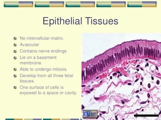
INSTRUCTIONS This exercise is set up so that the first thing you see is an image of a tissue without any explanatory text. Try to guess what type of tissue you are looking by identifying unique structures or characteristics you can see in the picture. After you have guessed, click again to get the answer and an explanation of characteristics you should be looking for. If you know which image you want to see, clicking on the name of the image in the index will take you directly to an image of that tissue.
Index: Non-epithelial tissues Quick Quiz Nervous Tissue Muscle Tissue Connective Tissue Loose connective t. Skeletal Muscle Multipolarneuron Cardiac Muscle Dense connective t. Smooth Muscle Blood Adiposetissue Hyaline cartilage Elastic cartilage Fibrocartilage Bone
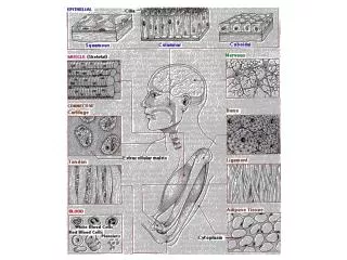
nuclei striations This is skeletal muscle tissue.Note themultiple nucleiat the edges of the muscle fibers. The striations are the overlapping patterns of muscle proteins in each fiber.
nuclei This is smooth muscle. Note that the nuclei are single, centrally located, and oblong. The shape of the nucleus mimics the shape of the cell it is in. Smooth muscle cells are described as “fusiform” in shape.
This is cardiac(heart) muscle. Note the centrally located nuclei, striations, and intercalated disks.
This is blood, which is classified as a connective tissue. Note that this image contains both red blood cells and white blood cells.
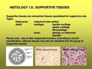
This is adipose (fat) tissue. Virtually all of this slide is filled with cells. (Single cell circled) Note that virtually all of the cytoplasm appears empty. This is where the fat is contained.
This slide is Connective tissue proper. More specifically, this is loose (areolar) connective tissue.
This tissue contains many collagen and elastin fibers, but relatively few fibroblasts (connective tissue cells). Fibers and fluid make up the extracellular matrix. Contrast this slide with the next slide.
This is also considered Connective tissue proper, but this tissue is dense connective tissue. Like areolar tissue, it contains few cells and a lot of fibers. The main difference is that they are more densely packed here.
This is cartilage, which is characterized by cells located in “little lakes” called lacunae (several are circled) within a relatively hard, glassy matrix.
There are 3 different types of cartilage, but you will be able to recognize each as cartilage if you see these distinct looking lacunae.
The light colored tissue above is hyaline cartilage. Can you see the lacunae?
Three are circled in red above. The black dots are the nuclei of the cells within them. The matrix looks clear.
Note that there are visible fibers in the matrix of this tissue, but you can still see lacunae.
This is fibrocartilage. The fibers in the matrix are very fine, and the lacunae are difficult to see, but they are still there. Do you see them?
This is a typical ground bone slide. Each cylindrical structure you see here is called an osteon, which is the structural unit of compact bone.
The dark structures indicated are the spaces where the cells resided when this tissue was alive. Like cartilage, the “holes” that bone cells rest in are called lacunae, even though they are less obvious than the ones in cartilage.
This is Nervous tissue. The big, dark cell in the middle is a multipolar neuron. Note the cellular extensions which are characteristic of this cell type.
Most multipolar neurons have 1 axon and a variable number of dendrites.
It’s hard to see axons and dendrites in a real specimen, because they are obscured by surrounding neuroglial cells. Normally, though, you can see the beginnings of them.
Quick Quiz For each of the following slides, try to guess which tissue you are looking at. Before you check the answer, be able to justify to yourself why you think it is a specific tissue.
Answer: Nervous Tissue (Large cell is a multipolar neuron.)