Structure of Epithelial Tissues
240 likes | 465 Vues
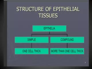
Structure of Epithelial Tissues. EPITHELIA. SIMPLE. COMPOUND. MORE THAN ONE CELL THICK. ONE CELL THICK. SQUAMOUS. CUBOIDAL. COLUMNAR. Feature of Epithelial Tissue. Closely packed cells with little extracellular material

Structure of Epithelial Tissues
E N D
Presentation Transcript
Structure of Epithelial Tissues EPITHELIA SIMPLE COMPOUND MORE THAN ONE CELL THICK ONE CELL THICK
SQUAMOUS CUBOIDAL COLUMNAR
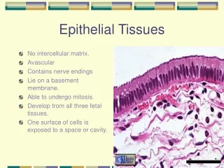
Feature of Epithelial Tissue • Closely packed cells with little extracellular material • Epithelial cells are arranged in continuous sheets, in single or multiple layers • Epithelial cells have an apical (free) surface, which is exposed to body cavity, lining of internal organ or exterior of body, and a basal surface which is attached to a basement membrane
Epithelia are avascular, blood vessels are located in nearby connective tissue; exchange of materials occurs by diffusion. Epithelia adhere firmly to nearby connective tissue by means of the basement membrane . Epithelia have a high capacity for renewal (high mitotic rate) since it is subjected to wear and tear.
Epithelial functions include: protection, filtration, lubrication, secretion, digestion, absorption, excretion, transportation, sensory reception, and reproduction
Covering and Lining Epithelium Epithelial layers are arranged as : simple epithelium -(one layer), where diffusion, filtration, secretion and absorption occur. stratified epithelium (several layers), protects underlying tissue from wear pseudo stratified epithelium (one layer that appears as several) because nuclei at different level; not all cells reach the surface; those that do have cilia or secrete mucus.
Cell Shapes • squamous(flat) for diffusion • cuboidal (cubelike) - cells have about the same height and width, produce secretions and function in absorption • columnar (rectangular) - cells are taller than they are wide protect underlying tissue, may have cilia, secretion or absorption.
Nucleus of Epithelial Cells Nucleus –conforms to shape of cell squamous :flattened disk cuboidal : spherical columnar : elongate/ovoid
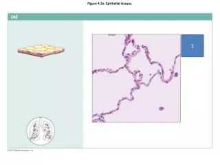
Simple Squamous & Cuboidal Epithelia • Squamous (single arrow) • Formed by flattened cells whose nuclei often appear to bulge outwards. • Found in places where there is movement of materials and even cells across the epithelium. Example here is from the loop of Henle in the kidney, also found lining all blood vessels, forming Bowman’s capsule in the renal cortex. • Cuboidal(double arrows) • In section cell profiles appear as squares with central nuclei. • Found lining tubules in kidney, walls of thyroid follicles Often involved in secretory functions
Nucleus of simple squamous cell Nucleus of simple cuboidal cell
Simple Columnar Epithelium • Found extensively in the gut, glands and ducts of glands. • Cells taller than they are wide although height variable. • Ovoid nuclei basally located. • Example here from the stomach. • Cells often involved in secretion (as in the stomach) and also in absorption as in the small intestine
Simple columnar epithelium comes in two forms: Nonciliated simple columnar epithelium - a single layer of nonciliated rectangular cells. Also functions in secretion and absorption. Specialized cells containing microvilli perform absorption. Goblet cells secrete mucus. Nonciliated columnar epithelia do not have cilia, and are found in the gastrointestinal tract and the gallbladder where they perform secretion and absorption
Ciliated simple columnar epithelium consists of a single layer of ciliated rectangular cells. Aids in movement. • Ciliated columnar epithelia • move mucus and other substances via cilia, and are found in the upper respiratory tract, the Fallopian tubes, the uterus, and the central part of the spinal cord.
Pseudostratified Columnar Epithelium The upper picture is from the seminal vesicle and the lower from the trachea. In both cases note how the nuclei are at different levels in the cells giving the appearance of more than one layer. In the case of the section of trachea the columnar cells carry a surface specialization (cilia - arrowed) and also have flask shaped goblet cells between them (G)