Download
1 / 33
340 likes | 609 Vues
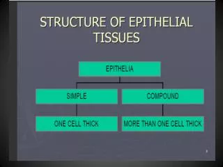
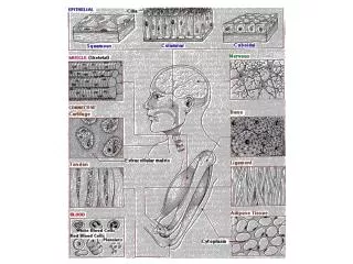
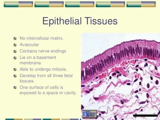
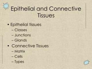
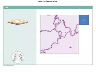
Figure 4.3a Epithelial tissues. 1. Figure 4.3b Epithelial tissues. 2. Figure 4.3c Epithelial tissues. 3. Figure 4.3d Epithelial tissues. 4. Figure 4.3e Epithelial tissues. 5. Figure 4.8a Connective tissues. 6. Figure 4.8b Connective tissues. 7. Figure 4.8c Connective tissues.

E N D
Figure 7.27a The humerus of the right arm and detailed views of articulation at the elbow. 15
Figure 7.4 Anatomy of the anterior and posterior aspects of the skull. 19 20
Figure 8.5d–f Movements allowed by synovial joints. 23 25 24
Figure 10.5 Superficial muscles of the body: Anterior view. 31 34 32 35 33
Figure 10.6 Superficial muscles of the body: Posterior view. 39 36 40 37 38
Figure 12.10a Midsagittal section of the brain. 44 41 42 45 43
Figure 12.26a Gross structure of the spinal cord, dorsal view. 46 47 48
Figure 12.26c Gross structure of the spinal cord, dorsal view. 49 50
Figure 15.24b Structure of the ear. 51 54 52 55 53
Figure 15.4a Internal structure of the eye (sagittal section). 56 59 57 58 60
Answer Key: • Simple squamous epithelium • Simple cuboidal epithelium • Simple columnar epithelium • Pseudostratified columnar epithelium • Stratified squamous epithelium • Nervous CT • Adipose CT • Reticular CT • Dense regular CT • Dense irregular CT • Lateral malleolus • Linea aspera • Pubis • Styloid process • Trochlea • Supraspinous fossa • Atlas • Hyoid • Optic canal • External occipital protuberance • Synarthrotic fibrous CT • Bursae • Flexion • Circumduction • Rotation • Inversion • Eversion • Amphiarthrotic cartilaginous joint • Amphiarthrotic cartilaginous joint • Rotation • Obicularis oculi • Pectoralis major • Tibialis anterior • Biceps brachii • Sartorius • Latissimusdorsi • Biceps femoris • Gastrocnemius • Deltoid • Gluteus maximus • Corpus calossum • Pituitary gland • Medulla oblongata • Pineal gland • Arbor vitae • Cervical enlargement • Lumbar enlargement • Caudaequina • Dorsal root • Ventral root • Malleus • Incus • Stapes • Semicircular canals • Cochlea • Lens • Aqueous humor • Cornea • Choroid • Retina