Download
1 / 34
340 likes | 560 Vues
Types of Epithelial Tissues in the Vertebrates. Outline. Tissue Types Epithelial Connective Muscular Nervous Organs Organ Systems Homeostasis Negative Feedback Positive Feedback. Levels of Organization. Tissue - Group of similar cells performing a similar function

E N D
Outline • Tissue Types • Epithelial • Connective • Muscular • Nervous • Organs • Organ Systems • Homeostasis • Negative Feedback • Positive Feedback
Levels of Organization • Tissue - Group of similar cells performing a similar function • Organ - Group of tissues performing a specialized function • Organ System - Collection of several organs functioning together • Organism - A collection of organ systems
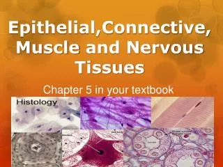
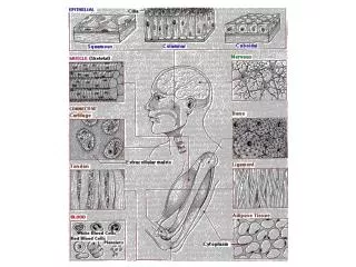
Types of Tissues • Four major vertebrate tissue types • Epithelial • Connective • Muscular • Nervous
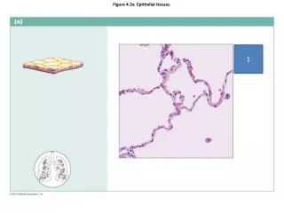
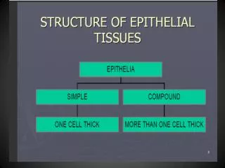
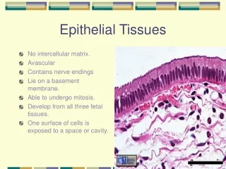
Epithelial Tissue • Epithelial tissue: • Forms a continuous layer over body surfaces • Lines inner cavities • Forms glands • Exocrine glands - Secrete products into ducts or cavities • Endocrine glands - Secrete products directly into the bloodstream • Covers abdominal organs • Three types of epithelial tissues: • Squamous – Flat cells • Cuboidal - Cube-shaped cells • Columnar – Pillar-shaped cells
Connective Tissue • Connective tissues consist of: • Fibroblast cells • A matrix containing collagen and elastic fibers • Loose fibrous connective tissue • Allows organs to expand • Dense fibrous connective tissue • Strong connective tissue • Tendons • Ligaments
Connective Tissue • Adipose Tissue • Insulates the body and provides padding • Cartilage • Classified according to type of collagen and elastic fibers found in the matrix • Cartilage cells (chondrocytes), lie in small chambers (lacunae) in the matrix
Connective Tissue • Compact Bone • Matrix is inorganic salts deposited around protein fibers • Bone cells (osteocytes) are located in lacunae • Lacunae arranged in concentric circles within osteons around tiny tubes (central canals)
Blood • Actually a connective tissue in which cells are embedded in a liquid matrix (plasma) • Red blood cells - erythrocytes • White blood cells - leukocytes • Transports nutrients and oxygen to cells • Removes carbon dioxide and other wastes
Muscular Tissue • Contractile cells containing actin and myosin filaments • Cells are called muscle fibers • Skeletal Muscle • Voluntary - Long, striated fibers • Smooth Muscle • Involuntary - No striations • Cardiac Muscle • Striated, but mostly involuntary • Bound by intercalated disks
Nervous Tissue • Nervous Tissue: neurons-specialized for the transmission of nerve impulses. • Made up of dendrites, a cell body, and an axon • Dendrites (like antennae) receive signals from sensory receptors. • Cell body contains the nucleus and organelles. • Axons conduct a nerve impulse away from the cell body. • Myelin-white matter • Unmyelinated fibers, cell bodies, and dendrites-grey matter • A nerve is a bundle of axons bound together by connective tissue. • Neuroglia support and nourish neurons
Nervous Tissue • Nervous system regulates body response to stimuli and has three main functions • Sensory input • Sensory (afferent) carrying impulses from the body to the CNS • Transmit info to the spinal cord • Data integration • Spinal cord and brain integrate • Decision is made regarding appropriate response • Motor output-(efferent) • Response is transmitted to effector (gland or muscle) • Effector initiates actual response
Neurons are not in direct contact with one another instead, a synapse separates them. Neurons communicate with other neurons by releasing chemical signals called neurotransmitters across a tiny gap.
Types of Neuroglial Cells • Astrocytes • CNS • mop up excess ions, etc • induce synapse formation • stimulate formation of tight junction between cells that make up the walls of capillaries. (Blood-brain barrier) • Schwann Cells • peripheral nervous system • myelinating cell • Oligodendrocytes • CNS • myelinating cell • Ependyma • CNS • ciliated • line central canal of spinal cord • line ventricles of brain • Regulate the production of CSF • Microglia • CNS • phagocytic cell
Integumentary system-Skin and derivatives • Functions of skin: Largest organ in the body • Covers and protects underlying body regions • Regulate body temperature • Contains sensory receptor- touch, pressure • Vital in maintaining homeostasis • Epidermis - Outer, thinner region • Stratified squamous epithelium-lacks blood vessels • As new cells enlarge, older cells are pushed outward, become keratinized, and are sloughed off • Melanocytes produce melanin (pigment) • Nails grow from specialized epidermal cells
Regions of Skin • Dermis - Deeper and thicker than epidermis • Fibrous connective tissue containing elastic and collagen fibers Contains: • Hair follicles • Sebaceous glands-secrete sebum • Receptors-specialized nerve endings • Nerve fibers • Blood vessels • Subcutaneous Layer - Loose connective tissue and adipose tissue located below dermis
Organ Systems • Body Cavities • Dorsal cavity / posterior (toward the back) • Contains the cranial cavity and the vertebral cavity • The brain is in the cranial cavity, and • The spinal cord is in the vertebral cavity • Ventral cavity/ anterior (toward the front) is divided by the diaphragm into • The thoracic cavity (includes heart and lungs) and • The abdominal cavity (most other internal organs) • The pelvic cavity
Homeostasis • The digestive system • Takes in and digests food • Provides nutrient molecules that re-place used nutrients • The respiratory system • Adds oxygen to the blood • Removes carbon dioxide The organ systems of the human body contribute to homeostasis
Homeostasis • The liver and the kidneys • Store excess glucose as glycogen-regulates blood glucose • Later, glycogen is broken down to replace the glucose used • The hormone insulin regulates blood glucose levels • The kidneys • Under hormonal control as they excrete wastes and salts • Regulate pH
Negative Feedback • Homeostatic Control • Partially controlled by hormones • Ultimately controlled by the nervous system • Negative Feedback is the primary homeostatic mechanism that keeps a variable close to a set point • Sensor detects change in the internal environment; • Regulatory Center activates an effector • Effector reverses the changes
Positive Feedback • During positive feedback, an event increases the likelihood of another event-a change occurs in one direction • Childbirth Process • Positive Feedback • Does not result in equilibrium • Does not occur as often as negative feedback
Review • Tissue Types • Epithelial • Connective • Muscular • Nervous • Organs • Organ Systems • Homeostasis • Negative Feedback • Positive Feedback