Download
1 / 17
200 likes | 629 Vues
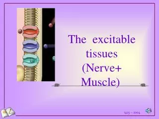
HISTOLOGY 1.9.: SUPPORTIVE TISSUES. Supportive tissues are connective tissues specialized for supportive role. Types: Embryonic: notochord (see earlier) Adult: cartilage: hyaline cartilage elastic cartilage fibrocartilage bone: spongy, or trabecular lamellar

E N D
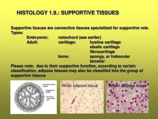
HISTOLOGY 1.9.: SUPPORTIVE TISSUES Supportive tissues are connective tissues specialized for supportive role. Types: Embryonic: notochord (see earlier) Adult: cartilage: hyaline cartilage elastic cartilage fibrocartilage bone: spongy, or trabecular lamellar Please note: due to their supportive function, according to certain classification, adipose tissues may also be classified into the group of supportive tissues White adipose tissue Brown adipose tissue
CARTILAGE Cartilage is a supportive tissue with firm ECM. The consistency of the tissue is hard, but bending. In the adult this tissue is normally avascular, aneural and alymphatic. It exists in the form of pieces of varied shape covered by the perichondrium. The perichondrium supplies the piece of cartilage with nutrients and oxygen via diffusion. Tissue components: cells (chondrocytes) ECM: fibers, ground substance Types: hyaline cartilage elastic cartilage fibrocartilage
Hyalin cartilage: Development of the hyaline cartilage: Clusters of mesenchymal cells chondroblasts chondrocytes (center of chondrification) (produce ground subst.) (in lacunae) Microscopic structure: Chondron (cell nests with isogenous cell groups) Pericellular matrix Territorial matrix Interterritorial matrix
Components of the ECM: Firm gel laced with collagen fibrils (same refraction index – non-visible) Ground substance contains GAGs: chondroitin sulphate, keratan sulphate, hyaluronic acid. Chondrocyte: lightly stained resting rounded cell LM TEM
Nutrition of the tissue: via diffusion from the perichondrium. Perichondrium:inner vascular, or cellular layer outer fibrous layer FPch: fibrous perichondrium ChL:cellular layer of perichondrium (appositional growth) ChB: chondroblast ChC:chondrocyte CN: chondron Lac: lacuna
Consequence of avascularity: low metabolic rate (bradtroph tissue) prone to degeneration (calcification, asbest degeneration) heals with persistent scar transplantation only with perichondrium Occurrence of hyalin cartilage: articulating surfaces of bones nose pharynx trachea bronchi appendicular and axial skeleton of embryo
Elastic cartilage: Occurrence: external ear epiglottis external auditory canal corniculate and cuneiform cartilages of the larynx Dense network of elastic fibers around the chondrons visible both with H.E. staining (Fig.1) and resorcin-fuchsin or orcein staining (Fig.2) 1 2
Fibrocartilage Occurrence: intervertebral disks menisci of stifle joints meeting of tendons and ligaments with the cartilage dog: between the atrial and ventricular heart muscles Collagen fiber bundle (rich in type I. collagen) Chondrocyte
Comparison of the three cartilage-types: Dominating tissue component: Physical property: 1. Hyaline cartilage: ground substance tensile strength chondrons with many cells 2. Elastic cartilage: elastic fibers elasticity chondrons with single cells 3. Fibrocartilage: collagen fibers great tensile strength very few chondrocytes against pulling forces 1 2 3
BONE Connective tissue with cells and fibers embedded in a hard non-bending ground substance. Functions: gives internal support for the body attachment for the muscles and tendons protects the brain and organs in the thoracic cavity contains the bone marrow main source of calcium for the organism Differences from the cartilage: canalicular system within the bone direct vascular supply appositional growth only It is a highly dynamic tissue that is renewed and remodeled throughout the whole life.
Structure of a long bone: Nutritive artery and vein
Tissue components of the bone: Cells: osteoblasts osteocytes osteoclasts Bone matrix – osteoid: ECM: organic: collagen fibers (flexibility) GAGs inorganic: hydroxyapatite crystals (firmness) (CA, CO3,PO4,OH, Na, Mg, Fe) Demineralized bone: looses firmness
The mature lamellar bone is built up by several osteons
Light microscopic structure of the lamellar bone Concentric lamellae (C): around Haversian canals (H) Interstitial lamellae (I): remnants of old osteons (space-filling) Circumferential lamellae (not shown): outer and inner surface of the bone V: Volkmann-canal Cross-section Osteon I Longitudinal section C V H H
Osteoblast: SEM TEM LM TEM Osteocyta: LM
Osteoclast LM TEM Osteoclast Bone matrix (osteoid) Howship-lacuna LM
Periosteum: Two layers: inner vascular and cellular osteogenic layer outer fibrous layer of collagen fibers and blood vessels Sharpey-fibers: collagen fibers continuous with the collagen fiber system of lamellae, fix the periosteum to the bone Endosteum: delicate layer of squamous cells lining the bone inside ion barrier plays role in the mineral homeostasis Bone marrow Endosteum Bone