Download

1 / 18
180 likes | 397 Vues
Learn about glomerular diseases, immune-mediated glomerulopathies, major clinical syndromes, and complications. Understand immune reactions, deposition of antibodies and complement, renal clinical syndromes, and complications of uremia.

E N D
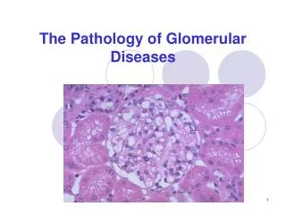
Significantly modified from “Serotonin” edition Overview of glomerular diseases Ali Al Khader, M.D. Faculty of Medicine Al-Balqa’ Applied University Email: ali.alkhader@bau.edu.jo
Contractile, capable of proliferation, makes ECM & releases mediators *Endothelial cells are fenestrated *Glomerular basement membrane (GBM) is mainly composed of: -Collagen IV -Laminin -Proteoglycans…polyanionic -Fibronectin …etc Elsevier. Kumar et al. Robbins basic pathology 9th…modified = filtration slit …bridged by thin slit diaphragm…mainly composed of: nephrin from adjacent foot processes Nephrin & its partner proteins (e.g., podocin) are very important in the selective permeability of the filtration barrier GBM: lamina densa inside & lamina rara on each side (interna & externa)
Kidney is the only or main organ involved = Glomerulopathies Kidney is involved additionally to other organs …also hypertension Elsevier. Kumar et al. Robbins basic pathology 9th…modified
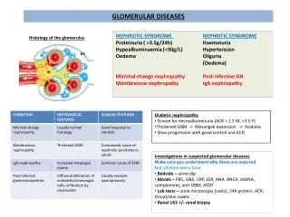
From the previous table…The following entities are the main immune-mediated glomerulopathies that you will hear about: • Anti-GBM antibody-mediated glomerulonephritis and Goodpasture syndrome • Postinfectious (or) Poststreptococcal glomerulonephritis • Membranous nephropathy • Mambranoproliferative glomerulonephritis I (MPGN I) & dense deposit disease • IgA nephropathy and Henoch-Schonlein purpura • Lupus nephritis
Regarding immune-mediated glomerulopathies (Glomerulonephritides) The antigen can be: -exogenous…e.g., streptococcal or antigens of other microbes, e.g., HBV, Plasmodium falciparum, Treponema pallidum or -endogenous…e.g., lupus nephritis or -unknown…e.g., Membranoproliferative GN (MPGN) Remember: membranous nephropathy Exogenous…e.g., endostroptosin in poststreptococcal glomerulonephritis Endogenous…e.g., lupus nephritis
Regarding immune-mediated glomerulopathies (Glomerulonephritides): We have an immune reaction with the following events occur at different quantities, patterns and combinations according to the disease we are talking about: -Leukocyte infiltration in the glomerulus -Antibody deposition in the glomerulus -Complement deposition in the glomerulus -Mesangial proliferation -Endothelial cell proliferation -Epithelial (visceral or parietal) cell proliferation -Fibrin deposition…remember that there is also a role of platelets and thrombin
Now we said that in immune-mediated glomerulopathies we may have deposition of immunoglobulins or complement… Where can we detect this deposition (mainly using immunofluorescence)?
You will hear about some names that are “Renal clinical syndromes” …which means that each of these syndromes is a group of symptoms/signs/lab results that points to a renal disease …each of these syndromes can be caused by more than one disease …The main 3 renal clinical syndromes associated with glomerular diseases are “Nephritic syndrome”, “Nephrotic syndrome” & “Rapidly progressive glomerulonephritis”
Other renal clinical syndromes • Asymptomatic hematuria, non-nephrotic proteinuria, or both: …mild glomerular abnormalities • Rapidly progressive glomerulonephritis (RPGN): …severe glomerular injury …loss of renal function in a few days or weeks …manifested by: -microscopic hematuria -dysmorphic RBCs & RBC casts -mild to moderate proteinuria …characterized microscopically by crescentic change in the glomeruli …remember that the microscopic counterpart of RPGN is: Crescentic glomerulonephritis …What is this?
Remember: -Increased urea and creatinine without symptoms = azotemia …with symptoms = uremia Other renal clinical syndromes, cont’d • Acute kidney injury (AKI)…previously called acute renal failure (ARF): …dominated by oliguria or anuria …recent onset of azotemia/uremia …can result from any acute condition affecting any part of the nephron(not always glomerular disease is the cause…remember that the most common cause of AKI is tubular) • Chronic kidney disease: …prolonged symptoms and signs of uremia …the result of progressive scarring in the kidney from any cause …may culminate in end-stage kidney disease …can result from any chronic condition affecting any part of the nephron
Complications of uremia Elsevier. Kumar et al. Robbins and Cotran pathologic basis of diseases 9th
Other renal clinical syndromes, cont’d These are not glomerular…
Immunofluorescence (IF) • When the nephrologist or pediatrician takes a kidney biopsy, he takes 3 samples (one fresh for IF, one in formalin for light microscopy, and one in glutaraldehyde for electron microscopy) • We take frozen sections of the kidney sample that was fresh and put them on slides • We use antibodies labelled by fluorochrome • The targets of these antibodies are IgG, IgA, IgM, C3, C4…etc. …for example: in one slide we direct anti-IgG to the tissue, in another one we direct anti-C3…and so on …if a disease for example is characterized by IgG deposition, the slide that we directed labelled anti-IgG to it will show positivity in the fluorescence microscope
IF, cont’d • Now the positivity is as the following: -Capillary(BM) VS Mesangial -Granular VS Linear
Granular VS Linear IF patterns Typical for: Anti-GBM GN Elsevier. Kumar et al. Robbins basic pathology 9th…(A, Courtesy of Dr. J. Kowalewska, Department of Pathology, University of Washington, Seattle, Washington.) *The deposition in these 2 examples is mainly a capillary deposition…not mesangial
A brief note on Anti-glomerular basement membrane antibody–mediated glomerulonephritis …α3 chain of the type IV collagen of the GBM is the target here …Sometimes the anti-GBM antibodies cross-react with basement membranes of lung alveoli, resulting in simultaneous lung and kidney lesions …this is called??? …less than 1% of GN cases but: Many instances are characterized by very severe glomerular damage with necrosis and crescents and the development of the clinical syndrome of rapidly progressive GN
A note to remember • Any renal disease (glomerular or nonglomerular) that destroys sufficient nephrons to reduce the GFR to 30-50% of normal…adaptive responses in the remaining renal tissue occur and this may be complicated by FSGS followed by global sclerosis entering a vicious cycle of nephron loss and maladaptation causing progressive glomerulosclerosis