Cellular Biology
Cellular Biology. School of Life Sciences Shaanxi Normal University. 1. CHAPTER 9 CELL PROLIFERATION AND REGULATION. The cell cycle can be described as 4 phases. I. Basic Concepts What is cell cycle:


Cellular Biology
E N D
Presentation Transcript
Cellular Biology School of Life Sciences Shaanxi Normal University 1
CHAPTER 9 CELL PROLIFERATION AND REGULATION
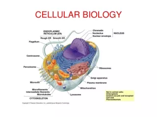
I. Basic Concepts What is cell cycle: A cell cycle means a cell proliferation procedures from the end of division to next end of division. The cell cycle can be described as 4 phases: ① G1 phase (gap1phase) means the gap time from the finished time point of mitosis to the beginning time point of DNA replication. ② S phase (synthesis phase) means the time when DNA is synthesized. H3-TDR can be inserted into new synthesized DNA in this phase only. ③ G2 phase (gap2 phase) means the gap time from the finished time point of DNA synthesis to the beginning time point of mitosis. ④ M phase or D phase (mitosis or division) means the time from the beginning to ending of mitosis.
The definition of cell cycle time: Percentage labeled mitoses (PLM) is a regular method for that. Label cells with pulse labeling method. Take cell samples at different time points. Display the labeled cells by autoradiography. Obtain the percentage of the labeled cells that proliferated. The terms for that are as the follows: TG1: Consisted time of G1. TG2: Consisted time of G2. TS: Consisted time of S. TM: Consisted time of M. TC: Consisted time of a cell cycle. PLM: Percentage of labeled mitoses. TDR: Thymidine. 3H or 14C is used to label TDR usually. All S cells were labeled by 3H S cells became M cells through G2 phase (The PLM is 0 in this time) The M cells appeared, the gap time between PLM = 0 and PLM > 0 is TG2 S cells turned to M cells, and PLM increased The peak means the late S cells turned to G1 cells through M phase (PLM = 100%) The gap time from M cell appeared to PLM = 100% is TM PLM decreased (The time from PLM appeared to decreased is TS) The time from PLM appeared to next PLM appeared is TC TC = TG1+TS+TG2+TM Obtain TG1 from this formula calculation.
Synchronization: Synchronization means all cells growing in a system are proliferating in same phase steps of cell cycle. The synchronization can be formed naturally or artificially. Natural synchronization: (No presentation, see your text book) Regulated (Artificial) synchronization: 1.Selective synchronization: (1) Mitosis selection: When the cultured cells grow to a logarithmic proliferation the mitosis cells are separated from dish bottom because the mitosis cell can not attach to the dish surface. If you shake the dish gently, M phase cells will be separated from dish surface. Collect the cell suspension, spin down and resuspend the cells with fresh medium again, continue to culture them. Repeat the steps above, you will get M phase cells. This method is simple, easy, and the cells kept no damaged. But you can not get M cells with 100% ratio. (2) Centrifuging isolation: The cells in different phases have different sedimentation coefficients. So, we can isolate each phase cells by centrifuge method. But the synchronized rate is not so good if use this method.
2.Regulated synchronization: • Blocking DNA synthesis: Use DNA synthesis inhibiting reagent to block DNA synthesis reversely, regulate all of cultured cells to be stopped at S phase or the G ending and S beginning. The reagents include 5-Fluoro-2'-deoxyuridine (FUDR), thymine deoxy-ribonucleoside (TDR), ADR, and GDR. • FUDR and TDR are better than others. For example, add excessive TDR into logarithmically growing cell culture (Hela: 2mol/L; CHO: 7.5mol/L), check cell growth each day to identify stopped culture growth. Wash cells and add fresh medium again and continue to the culture. When the time of H3 releasing is longer than TS, add excessive TDR again. All cells will be stopped at G1/S. • The advantage of this method is very high synchronization rate (almost 100%). The disadvantage of the method is that the size of some cells become bigger. • (2) Blocking mitosis metaphase: Damage micro-tubes and stop cell proliferation at mitosis metaphase. The reagents include colchicine and colchicinamide. Colchicinamide is better than colchicine because of its lower toxicity. • The growth of cells by this method is good. But it is difficult for the cells to be cultured reversely.
II. mitosis • The types of cell division: • Amitosis: Amitosis is also called as direct division. For amitosis, the nucleus become longer and a constriction is formed at middle of the long nucleus, then, the nucleus and plasma is separated into two new cells. No spindle is formed and no chromosome change can be found in amitosis. Amitosis can be observed in prokaryotic cells and some special eukaryotic cells, such as the cells of fetal membrane, muscle cells, and others. • Mitosis: Mitosis is also called as indirect division. The spindle and chromosome change can be observed in mitosis. Generated chromosomes can be separated into two new cells equally. Mitosis is the popular division style in advanced animal and plant cells. • Meiosis: In meiosis, the chromosomes are once replicated, but the cells twice proliferated. Meiosis is the division style for the germs of advanced animals and plants. • Mitosis: • For the convenience to describe, we divide mitosis as 6 stages: interphase, prophase, premetaphase, metaphase, anaphase, and telophase. The interphase include G1, S, and G2 phases in that the DNA replication is prepared. I introduce other phases involved with mitosis to you as the follows:
Prophase: In prophase, the following events happen: ① chromatin is condensed; ② set up polar sites and start to form spindle; ③ nucleolus is disassembled; ④ Nuclear membrane (envelope) disappeared. In prophase, the chromatin became short and visible under microscope. Each chromosome include two chromatids. Two centrioles have already formed in S phase, and they move to polar sites in prophase. The spindle microtubes will be formed between the both centrioles. Spindle is formed, and nuclear membrane disassembled. The spindle microtubes include follows: ① Kinetochore mt. ② Astral mt. ③ Polar mt or overlap mt. The spindle microtubes play roles to link polar bodies and separate chromosomes and division bodies. There two types of motor protein are involved in these changes: dynein and kinesin.
Premetaphase: Premetaphase means the stage from the membrane disassembly to chromosomes were managed to equatorial plane. Spindle microtubes extend into center and combine to kinetochore. Left: Premetaphase; Right: Metaphase (From http://www.wadsworth.org)
Metaphase: Metaphase means the stage from the chromosomes managed on equatorial plane to the paired chromatids separated to polar sites. Left: Metaphase; Right: The microtubes linked to chromosomes (http://www.wadsworth.org)
Anaphase: In anaphase, paired chromotids are separated and move to polar sites. The paired chromotids are separated in anaphase (http://www.wadsworth.org)
Anaphase can be described as two stages: ① Anaphase A means the stage when the chromosomes are moving to the centrosomes (polar sites). ② Anaphase B means the stage when the distance between both polar sites is becoming longer. The changes in both stages above are carried out by the cooperation of microtubes and motor molecules. The anaphases A and B were identified out by using taxol (a reagent) or others. Taxol can inhibit anaphase A. There is no anaphase B in plant cells.
Chromosomes are separating off in anaphase A, and the both polar sites are moving away each other in anaphaseB
Chromosomes are separated away by the cooperation of motor protein and microtubes system
Telophase: Telophase means the stage from the time point when the new chromosomes moved to polar sites to the time point when two new cells have been formed. Telophase includes new nuclei generation stage and cell plasma division (cytokinesis) stage.
New nuclei generation: New nuclei generation takes an opposite way to the one taken in prophase: Chromosome chromatin appearance of nucleolus envelope formation. Nucleolus is formed by the nucleolus organization regions (NORs) of chromosomes. Plasma division (cytokinesis): For the eggs of insects, nucleus can be cleaved many times without cytokinesis. The cytokinesis of animal cells is started by forming a contraction ring on plasma membrane. If the cells are treated by cytochalasin, or antibodies against myosin or actin, the contraction ring will not be formed. This indicates that the contraction ring acts in a mechanism like muscle contraction.
The plasma cleavage of plant cell is different from animal cell. The microtubes located in polar sites will disappeared in anaphase or telophase. But the microtubes located in center will be remained and generated. So, the phragmoplast will be formed. The vesicles from Golgi body will be transported into the phragmoplast to form the cell plate and the filament between cells. The vesicles from Golgi bodies are fused to cell plate to form cell wall, then, the cell will be separated as two new cells.
III. Meiosis • During meiosis, the cell is cleaved twice, but the DNA is replicated once only. So, the number of chromosome for new generated cell is decreased to 23 for each cell (human germ cell). So, we say that human germ cells are haploid cells (n = 23). They will return back to diploid (2n = 46) by the fertilization with 23 chromosomes from father and other 23 chromosomes from mother. There is an exchange between homologous chromosomes by that the genetic information for new generation will be variegated, the evolution development for species will be enhanced, and the number of chromosome for cell will be kept no changed consistently. • Meiosis can be sorted as 3 major types: • Gametic meiosis (terminal meiosis). The meiosis is combined with germ cell development together. For example, one spermatocyte can be cleaved as four spermatids through meiosis. The spermatid will be developed as sperm. One ovocyte (oocyte) can be cleaved as one ovum and 2 or 3 polar bodies. • Sporic meiosis (intermediate meiosis). Sporic meiosis is the type of meiosis for plants. Sporic meiosis and germ cell development are different procedures and not combined together. The gametocytes (sporocysts) will be developed as haploid microspores and macrospores in sporic meiosis. Microspore will be developed as microgamete (male gamete), and macrospore will be developed as macrogamete (female gamete).
(3) Zygotic meiosis (initial meiosis). Zygotic meiosis is the meiosis type for fungi and some bacteria. The meiosis happens after the generation of zygote and will form the haploid sporozoite. Somatic meiosis can be found in some living things, such as mosquito larva. Meiosis is composed of both continued cleavages (Meiosis I and II). Homologous chromosomes will be separated away in meiosis I usually. So, we can also call it as heterotypic division or reductional division. Paired chromosomes will be separated away in meiosis II like mitosis. We can also call it as homotypic division or equational division.
Like mitosis, the meiosis can be divided as several stages for the convenience to describe it. Premeiotic interphase (premeiosis): Premeiosis includes G1, S and G2 phases. Mitosis turns to meiosis in G2 phase. Meiosis: 1. Meiosis I: Premeiosis I: The main steps of meiosis are carried out in premeiosis I. For the convenience of statement, we can divide the premeiosis as 5 stages as the follows: (1) Leptotene: Chromosomes appear as filaments with beaded chromomeres. The chromosomes have replicated at this time, but the paired chromatids can not be observed under microscope yet. This stage is also called as synizesis because the chromatids are overlapped together. For some species, the chromatid filament is linked to the nuclear membrane with one terminal, and another terminal extends into nuclear plasma. So, some people call it as bouquet stage. (2) Zygotene: The homologous chromosomes will be paired in this stage, that is called synapsis. The homologous chromosomes will form the synaptonemal complex (SC). We can see the paired chromosomes combined together under optic microscope and call it as bivalent. Each pair of chromosomes is replicated and contains 4 chromatids called as tetrad.
(3) Pachytene: This stage can go on for several days. Chromosomes will become short and combine closely each other. The homologous chromosomes unpaired can be exchanged partially each other at this time. (4) Diplotene: The linked homologous chromosomes of SC start to separate each other, and keep a junction at chiasma. SC is disappeared in this stage. The chiasma can move to the terminal of the bivalent. We call this movement as terminalization. The diplotene is much longer in animal germ cells than in plant germ cells. Human ovocytes have turned to diplotene in 5 months fetus, but they will stay in this stage till to be exported (12 – 50 years old woman). (5) Diakinesis: The bivalents become short, move to the peripheral area of nucleus, and distribute to every where in nucleus. So, this stage is the best time to observe chromosomes in meiosis cells! The shape of bivalents can be V or O at this time. Nucleolus will disappear at this time. But some plants, such as corn, keep nucleolus visible still at this time.
Metameiosis I (Metaphase I): Nucleolus disappeared and nuclear membrane disassembled indicate that the cell has turned to metameiosis I. In this stage, chromosomes are arranged on the equatorial plane. Each bivalent has 4 centromeres, and the centromeres of paired chromatids are located to the polar side of spindle. We call this localization as co-orientation. Anameiosis I: The homologous chromosomes of bivalent are separated and move to both polar sites. The number of chromosomes in each polar area is decreased by half because the separation of homologous chromosomes. The separation and distribution of homologous paired chromosomes in both polar sites is absolutely randomly to make the recombination of the chromosomes from mother and father, that is very beneficial to the genome mutation. There are 23 pairs of chromosomes in a human cell with 223 recombination types. So, excepting monozygotic twins, it is almost impossible to have the generations with same genetic features. Telomeiosis I: After chromosomes move to polar areas, they are despiralized, and the nuclear membrane and nucleolus will be assembled again. Meantime, the plasma will be cleaved.
Intermeiosis: It is a short stage between meiosis I and meiosis II without DNA synthesis. 2. Meiosis II: It is similar to mitosis, and can be divided as 4 stages: premeiosis II, metameiosis II, anameiosis II, and telomeiosis II. By the meiosis, a spermatocyte can form 4 sperms, and a ovocyte can form 1 ovum and 2 or 3 polar bodies. Synaptonemal complex (SC): Synaptonemal complex (SC) is formed by two homologous chromosomes in the zygotene. SC is associated with the pairing, exchanging, and separating of homologous chromosomes. The both sides of SC are 40nm lateral element, and a 100nm intermediate space is located in center. A 30nm central element is located in center of intermediate space. The 7 – 10nm SC fibers are horizontally arranged between lateral element and central element, that is make SC looked like a ladder. The spherical recombination nodules (RNs) can be observed in the SC stained by phosphotungstic acid. RNs are the sites where the homologous chromosomes are crossed. Some enzymes for gene exchange are located on RNs.
SC fibers Lateral element Intermediate space Lateral element A SC of an insect
IV. Regulation of Cell Cycle The background of research on the regulation: Rao and Johnson (1970、1972、1974) synchronized cultured Hela cells at different phases, then, mixed them with M phase cells to induce the cell fusion intermediated by inactive Sendai virus. They found that the interphase cells of the fused cells can form the different shapes of prematurely condensed chromosome (PCC). They named this course as premature chromosome condensation. The PCC of G1 phase is leptotene because the DNA is not replicated yet. The PCC of S phase is like powder because the DNA has been replicated on many sites. The PCC of G2 phase is diplotene because the DNA replication has been finished.
The described above just shows you that the fused M phase cells of same type of cells can introduce PCC. Actually, the fusion of different types of cells can introduce PCC also. For example, the fused M phase cells of human and toad can introduce PCC also. The result above indicates that M phase cells can secret some factor that can promote the proliferation of interphase cells. The factor is called as maturation promoting factor (MPF). In 1960s, Yoshio Masui found the extract from the matured frog egg can promote the Germinal Vesicle Breakdown (GVBD) of immature frog egg. Sunkara injected the extract from different phase Hela cells into frog egg, they found that the extract from G1 and S phase cells can not introduce GVBD, but the extract from G2 and M cells can promote GVBD. They named this extract as mitosis factor (MF). The same factors were found in many other types of cells later on. All of they are called as MPF. In 1960s, Leland Hartwell isolated tens cell division cycle gene (CDC) from some yeast cells. For example, the cdc28 gene plays an important role on the G2/S exchange. By the research on the sensitivity of yeast to radioactive rays, Hartwell put forward a new concept, checkpoint, means that a cell cycle will be stopped when the DNA is damaged.
In 1970s, Paul Nurse, et al, found many cell cycle regulatory genes from yeast. For examples, the mutants of cdc2, cdc25 can not take mitosis at regulated temperature, the mutant of wee1 can start division early, and the mutant cdc25 and wee1 can take division normally. By the further experiments, they found cdc2 and cdc28 encode a 34KD protein kinase to promote cell cycle. The genes wee1 and cdc25 can inhibit or enhance cdc2. That is why the mutant of cdc25 and wee1 can take normal division. In 1983, Timothy Hunt found some special protein from the fertilized egg of sea urchin firstly. The level of the protein in the each phase of the egg cell cycle is changed obviously. This protein synthesis can be started at G1, increased to a peak at G2/M, and disappear after M phase. He named the protein as cyclin. Cyclin was lately found in other animals and yeast. The cyclin mRNAs from other animals can promote the maturation (division) of the fertilized frog egg. Because the great contribution to the cell biological development, all persons above became Nobelist later.
If the expression of Cdc25 is inefficient, the cell grows long without division; If the expression of Wee1 is inefficient, the cell starts division early (Cell is small)
In 1988, M. J. Lohka purified the MPF from toad, and found that the MPF is composed of 32KD and 45KD proteins, the complex of the both proteins can phosphorylate many other proteins. Paul Nurse(1990)found that the 32KD and 45KD proteins are the homologous molecules for cdc2 and cyclin B. This finding combines the main research projects above together. On Oct. 8, 2001, American, Leland Hartwell, British, Paul Nurse and Timothy Hunt won the Nobel prize because of their great contribution to the researches on the cell cycle regulation.
34KD protein kinase MPF = cdc2 + Cyclin B
Cyclin-dependent kinase (CDK): Cdc2 can get kinase activity when it combines to cyclin, so, cdc2 is called as cyclin-dependent kinase (CDK) or CDK1. Activated CDK can phosphorylate proteins to take functions. For example, it can phosphorylate lamina protein to disassemble nuclear skeleton and envelope. By this disassembly, the cell cycle can be kept for continued division. So far, 7 CDKs (CDK1 – CDK7) have been found in animals. Each of them contains a similar kinaselike domain with a conserved peptide sequence, such as PSTAIRE, that is the binding site to cyclin. CDK inhibitor (CDKI): There are CDK inhibitors (CDKI) in cells that can regulate cell cycle negatively (inhibiting). Two families of CDKI have been found so far: ① Ink4 (Inhibitor of cdk4), such as, P16ink4a, P15ink4b, P18ink4c, and P19ink4d. These Ink4s can inhibit the complex of cdk4·cyclin D1 and cdk6·cyclin D1 specifically. ② Kip (Kinase inhibition protein), such as, P21cip1 (cyclin inhibition protein 1), P27kip1(kinase inhibition protein 1) and P57kip2 that inhibit the kinase activity of most of CDKs. P21cip1 can combine the helper factor of DNA polymerase, proliferating cell nuclear antigen (PCNA), to inhibit DNA synthesis.
Cyclin: Cyclin can both activate CDK and manage what substrate will be phosphorylated where and when to promote cell cycle. So far, more than 30 cyclins have been isolated from yeast and animals. The cyclins in vertebrates include A1-2, B1-3 , C, D1-3, E1-2, F, G, and H. All of them can be sorted as 4 types: G1, G1/S, S, and M. Each type of cyclin contains an about 100aa (amino acid) conserved sequence called as cyclin frame that can intermediate the combination of cyclin and CDK. Cells will express G1 phase cyclin D under the growth factor activation. The cyclin D can combine CDK4 and CDK6 to phosphorylate the downstream proteins, such as Rb, and the phosphorylated Rb can release out the transcription factor, E2F, to promote many transcriptions of genes, such as, the genes of cyclin E, cyclin A, and CDK1. Cyclins
The combination of Cyclin D and CDK promotes Rb to release out the combined transcription factor, E2F
In G1/S, cyclin E combines CDK2 to promote the cell turn to S phase through G1/S restriction point. If you use antibody against Cyclin E to the cultured cells, your cells will be stopped at G1. If you use antibody against Cyclin A, the DNA synthesis in your cells will be inhibited. In G2/M, cyclin A and cyclin B can combine CDK1 to phosphorylate substrate protein to cause the downstream events. For examples, phosphorylation of histone H1 can cause the condensation of chromosomes, and phosphorylation of lamina proteins can cause the disassembly of envelope. The cyclic changes of cyclin
In the metaphase with peak of MPF activity, by an unknown pathway, anaphase promoting complex (APC), can be activated to combine ubiquitin to cyclin B, and cause cyclin B degenerated by proteasome. A cell cycle is finished.
There is a sequence associated with cyclin destruction on the N terminal of mitosis phase cyclin. We call this sequence as destruction box. At the peak of MPF, the ubiquitin ligase can promote the combination of ubiquitin and cyclin. The ubiquitin combined cyclin can be hydrolyzed by 26S proteasome. G1 phase cyclin can be hydrolyzed by same way, but there is no destruction box on it. There is a PEST sequence on the C terminal of it that is associated with its destruction. Ubiquitin is composed of 76 amino acids, it is highly conserved, and exists in all eukaryotic cells. That is why it is called as “Ubiquitin”. Ubiquitin functions like a marker to be destructed. All proteins combined ubiquitin can be recognized and destructed by proteasome. It is the popular way by that short life proteins and abnormal proteins in cells can be cleaned. In the polyubiquitination, E1, an ubiquitin-activating enzyme, gets energy by hydrolyzing ATP to activate ubiquitin, then, E1 transfers the activated ubiquitin to E2, an ubiquitin-conjugating enzyme. Finally, E3, an ubiquitin-ligase, links the ubiquitin to target protein. There are two types of ubiquitin ligases at least involved with cell cycle regulation: skp1-cullin-F-box protein (SCF), a complex composed of 3 proteins, links ubiquitin to G1/S cyclin and some CKIs, APC will combine the ubiquitin to M phase cyclin like what is shown by the fig above.
DNA replication should be once carried out, andis once carried out exactly in a cell cycle: DNA replication is started from the origins of replication that are the autonomously replicating DNA sequences (ARSs) distributed in chromosome, and I presented them to you in last chapter. In the cell cycle, these DNA replication origins are combined with origin recognition complex (ORC) that can be bound by other regulatory factors. Cdc6 is the one of these factors. Another very important factor is from a special protein family named minichromosome maintenance protein (MCM). MCM is the licensing factor to the DNA replication, and 6 MCMs (MCM2 – MCM7) have been found. If any one of MCMs is absented, the DNA replication will be stopped. In G1 phase, the level of cdc6 is increased quickly, and it is combined to ORC to promote other proteins including MCM binding to ORC, and form the pre-replicative complex (pre-RC). MCM is DNA helicase actually. S phase CDK (S-CDK) both triggers pre-RC to start DNA replication and blocks the DNA re-replication because S-CDK can phosphorylate cdc6 to separate it from ORC. Phosphorylated cdc6 can be degenerated by the SCF joined polyubiquitination pathway. S-CDK can also phosphorylate MCM to inactivate it. Other CDKs can also block the pre-RC secondary formation. By all pathways described above, it is ensured that the DNA replication should be carried out once only, and is carried out once only in a cell cycle.
Each cell cycle triggers DNA replication once only (from Molecular Biology of the Cell 4th ed.)