Download
1 / 109
1.09k likes | 1.15k Vues
1.1.4-1.1.12; 1.2.1 – 1.3.5; 1.4.7. CELLULAR BIOLOGY. Chapter 6: Cell Structure. Microscopy: Invention and improvement in 17 th century led to the discovery of cells. A) Review of the history 1590 Janson: Invented compound microscope

E N D
1.1.4-1.1.12; 1.2.1 – 1.3.5; 1.4.7 CELLULAR BIOLOGY
Chapter 6: Cell Structure • Microscopy: Invention and improvement in 17th century led to the discovery of cells. A) Review of the history 1590 Janson: Invented compound microscope 1665 Robert Hooke: coined term “cell”, examined cork with improved scope
More History 1650-1700 Anton von Leewenhoek: observes first living cells; unicellular with nuclei; called animalcules 1838 Mathias Schleiden: all plants made of cells or their derivatives 1839 Theodor Schwann: worked with Schleiden and proposed cell theory which suggested that all animals and plants were made of cells, and that within an organism these cells are identical.
History continued • Purkinje: fibers that transmit stimulus to myocardial cells of ventricles of heart bear his name; coined term “protoplasm” for the living content of cells. 1855: Rudolf Virchow: studied pathogenic organisms and showed that “omnis cellula e cellula” – all cells arise from preexisting cells (by cell division).
Summary of cell theory: • 1. Living organisms are composed of cells (evidence: work of Schleiden and Schwann and early microscopists). • Exceptions: Skeletal muscle fibers which contain hundreds of nuclei. Fungi which consist of thread-like structures (hyphae) which contain many nuclei. Many tissues, such as bone, contain a greater volume of extracellular material. • 2. Cells are the smallest unit of life. Evidence: cannot grow pieces of cells (organelles) and have them survive. • 3. Cells come from pre-existing cells. Evidence: Work of Francisco Redi and spontaneous generation.
Unicellular (or Acellular) Organisms: • Carry out all of the functions of life which include: a) metabolism b) response c) homeostasis d) growth e) reproduction f) nutrition
B. Technology • Light microscope (from Renaissance): 2 great advantages over the unaided eye. • Increased magnification: how much larger an object is made to appear compared to its real size. Limit = 1000x • Increased resolution: minimum distance between 2 points that can still be distinguished as 2 points. Limitation: wavelength of visible light. Max. resolution = 0.2 um. Size of small bacteria.
Other advantages of light microscope • Increased contrast: makes details that can be resolved stand out better to the eye. • Advantage over next generation of microscopes: can watch movement/living organisms • Can see color (advantage over electron microscope).
Electron Microscopes (1950s) Advantage over light microscope: • Increased resolution: uses shorter wavelengths – beam of electrons. In comparison to light microscope = 1000x resolving power. 0.2 nm (1 nm = 1/1000um). Or 2x diameter of a H atom. • Increased magnification: allows clear identification of subcellular organelles; used to study ultrastructure = cell’s anatomy as resolved by electron microscope. Definition of organelle: one of several formed bodies with a specialized function, suspended in cytoplasm and found in eukaryotic cells.
2 types of electron microscopes • Transmission e.m.: internal structures of cell can be seen • Scanning e.m.: study surface of specimen; get 3-D images. Both have improved our understanding of many biological and physical phenomena (e.g. viral penetration).
II. Measurements of cells and organelles 1 cm = 10-2 meter (0.4 inch) 1mm = 10-3 meter 1 micrometer (um) = 10-3 mm 1 nanometer (nm) = 10-3 um Atom = .1 nm Molecule = 1 nm Thickness of membranes = 10 nm
More measurements Viruses = 100 nm Bacteria = 1 um Organelles = up to 10 um cells = up to 100 um Chick egg = 5 cm Nerve/muscle cells in leg = 1 m Human height > 1 m
III. Size of cells: Why so small? The larger the cell, the harder it is to communicate from one part to the other; more difficult to move substances through diffusion. As cell size increases, volume increases. Chemical reaction rate is a function of volume. Transport is a function of surface area. Take this exercise:
Increasing the cell radius by 10 Cell Radius (r) 1 cm 10 cm Surface area 12.57 cm2 1257 cm2 4 p r2 Volume 4.189 cm34189 cm3 4/3 p r3
So, increasing a cell’s radius by a factor of 10, increases the surface area by 100 and the volume by a factor of 1000.
Surface Area to volume ratio • The surface area to volume ratio is a factor that limits cell size. Rate of metabolism of a cell is a function of its mass/volume; whereas the rate of exchange of materials and energy (heat) is a function of its surface area. • By dividing a large cell into many smaller cells, we restore surface area-to-volume ratio for acquiring nutrients and expelling wastes.
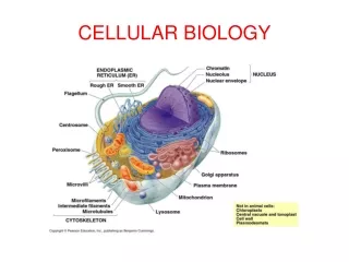
IV. Features that all cells have in common • Plasma membrane: encloses a cell and separates its contents from its surroundings. Composed of: phospholipid bilayer with proteins embedded. Proteins: 1) transport proteins help molecules and ions move across the plasma membrane (pumps and channels). 2) receptor proteins which induce changes within the cell when they come in contact with specific molecules in the environment (hormones). 3) markerswhich identify the cell as a particular type (important in multicellular organisms whose cells must be able to recognize one another as they form tissues.
B. Nucleoid or nucleus Nucleoid or nucleus contains genetic material. Prokaryotes (bacteria): most DNA resides near center of the cell in area called nucleoid. Eukaryotes: DNA segregated in a nucleus. C. Cytoplasm: semifluid matrix of sugars, amino acids, proteins that the cell uses to carry out everyday activities. In eukaryotes also contains specialized membrane bound compartments called organelles.
V. Prokaryotic vs. Eukaryotic • Distinguished by structural organization. • Prokaryotes (Monerans): bacteria and cyanobacteria. Be able to draw a generalized prokaryotic cell as seen in electron micrographs. They lack membrane-enclosed organelles. DNA is not associated with protein, chromosome is frequently circular and double stranded; hereditary material is located in nucleoid region; no membrane separates DNA from the rest of the cell.
More bacteria: Plasmids • Many bacterial cells also possess small, independently-replicating circles of DNA called plasmids. They contain only a few genes, usually not essential for the cell’s survival. R plasmids contain antibiotic resistance genes.
Bacterial Ribosomes • Bacteria have a large number of ribosomes. These ribosomes are smaller than the ribosomes of eukaryotic ribosomes. • 70s vs. 80s where s is a svedberg unit, which is a unit of sedimentation (where it centrifuges to in a salt gradient). • Since protein synthesis machinery is different in prokaryotes, bacterial ribosomes can be targeted by antibiotics (tetracycline).
Bacterial Plasma Membrane Modification • Plasma membrane: sometimes folds in to form a mesosome. Sometimes this is extensive. Functions in cell respiration or photosynthesis. • Outside plasma membrane: rigid cell wall and an outer jellylike slime capsule. Capsule is sticky and is still another protective layer. Enables the organism to adhere to their substrate and provide additional protection. Peptidoglycan present in cell wall (not present in Eukaryotes).
Other features • Flagella (locomotion): long, thread-like structures protruding from the surface of a cell that are used in locomotion and feeding. Protein fibers; 1 or more per cell. • Pili(adherence)
B. Eukaryotic Cells • More complex cells that are compartmentalized. • Internal membranes partition the cell; many enzymes built into the membrane. • Can perform many functions simultaneouslyin different parts of the cell, independent of one another.
Plant Cells • Often have a large membrane-bounded sac called a central vacuole which stores proteins, pigments and waste materials. • The membrane around this central vacuole is called the tonoplast.
All Eukaryotic Cells • Both plant and animal cells contain vesicles, smaller sacs that store and transport a variety of materials. • Inside nucleus, DNA is wound tightly around proteins and packaged into compact units called chromosomes. • All eukaryotic cells supported by an internal protein scaffold, the cytoskeleton. • Cells of fungi, plants, and many protists have cell walls composed of cellulose myofibrils or chitin fibers (fungi) embedded in a matrix.
Stem Cells • Cells that retain the capacity to divide and have the ability to differentiate along different pathways. • E.g. Use: in 2005, stem cells were used to restore the insulation tissue of neurons in laboratory rats, resulting in subsequent improvements in their mobility. • E.g. Use: blood stem cells taken from human umbilical cords and placentas can be harvested, tissue typed, stored, to be used with a human match following bone marrow destruction by chemotherapy in the treatment of leukemia.
VI. Tour of Eukaryotic Cell • Nucleus: • largest and most easily seen organelle. Often in center of cell. • Contains most of the genetic information • Averages 5 um diameter
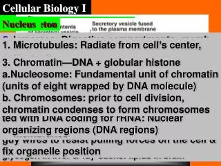
Nucleus and Nuclear envelope • Nucleus is enclosed by a nuclear envelope a) double membrane: 2 phospholipid bilayer membranes; outer membrane is continuous with cytoplasm’s interior membrane system (endoplasmic reticulum). b) On nuclear side, a network of protein filaments (the nuclear lamina) which stabilizes nuclear shape.
c) Perforated by pores: shallow depressions about 100 nm in diameter, 50-80 nm apart where the two membrane layers pinch together. Filled with proteins that act as molecular channels. Proteins move in (DNA polymerase); RNA and protein-RNA complexes formed in the nucleus, exported to the cytoplasm. d) Chromatin: complex of DNA and packaging proteins called histones; when DNA gets ready to divide, DNA coils up around histones in a highly condensed form called nucleosomes. Uncoiled chromosomes let the enzyme, RNA polymerase, make RNA copies of DNA to be translated into proteins.
Nucleolus e) nucleolus: spherical region in nucleus of nondividing cells; consists of nucleolar organizers (specialized regions of some chromosomes) with multiple copies of genes for rRNA; site of genes for rRNA synthesis; functions to assemble ribosomes.
VII. Ribosomes • Site of protein synthesis • 60% rRNA and 40% proteins • Two subunits: small and large • Prokaryotic cells may have a few 1000 ribosomes, a metabolically active eukaryotic cell (e.g. liver) may contain several million. • Proteins that function in cytoplasm are made by free ribosomes suspended in cytoplasm. • Proteins bound within membranes or destined for export from the cell are assembled by ribosomes bound to the endoplasmic reticulum.
Endomembrane System Definition: membranes interrelated directly through physical contact or indirectly through vesicles. Vesicles: membrane enclosed sacs that are pinched off portions of membranes moving from one membrane to another. Endomembrane system includes: nuclear envelope, endoplasmic reticulum, golgi apparatus, lysosomes, vacuoles, plasma membrane
XI. Endoplasmic Reticulum (ER) • Network of internal membranes; forms compartments and vesicles; participates in protein and lipid synthesis. • Largest of the internal membranes • Composed of lipid bilayer embedded with proteins; creates series of channels and interconnections between its folds. • Part of the endomembrane system
a) Smooth E.R.: cytoplasmic surface lacks ribosomes • Synthesizes lipids, phospholipids and steroids e.g. testes, ovaries and skin oil glands are rich in smooth e.r. • Participates in carbohydratemetabolism e.g. smooth e.r. in liver cells contain an embedded enzyme that removes phosphate from the first product in the hydrolysis of glycogen. • Detoxifies drugs and poisons. E.g. in liver enzymes add –OH groups to drugs and poisons made them soluble in cytosol so that they can be excreted from the body. • Stores calcium ions necessary for muscle contraction.