Download

1 / 37
370 likes | 387 Vues
Learn about the importance, procedures, and treatment of screening for retinopathy and nephropathy, two common complications of diabetes. Understand the screening pathway, classification of retinopathy, and the prevalence of diabetic nephropathy.

E N D
SCREENING FOR RETINOPATHY & NEPHROPATHY Prof.V.Mohan.,M.D.,Ph.D.,D.Sc. DIRECTOR M.V.DIABETES SPECIALITIES CENTRE, VISITING PROFESSOR OF DIABETOLOGY SRI RAMCHANDRA MEDICAL COLLEGE, PORUR PRESIDENT MADRAS DIABETES RESEARCH FOUNDATION, CHENNAI PROFESSOR OF INTERNATIONAL HEALTH UNIVERSITY OF MINNESOTA, USA 1
CARDINAL PRINCIPLES FOR SCREENING (WHO) 1. Important health problem with a presymptomatic state 2. Acceptable screening procedures (both by public and health care professional) 3. Safe, effective and universally agreed treatment 4. Economic cost of screening and treatment should be less than that for diagnosis and treatment
THE SCREENING PATHWAY Healthy Disease or precursor detectable Screening possible Symptoms develop Intervention to avert disease development or its consequence Advanced disease Life prolonged Death
DIABETIC RETINOPATHY CLASSIFICATION NON - PROLIFERATIVE DIABETIC RETINOPATHY PROLIFERATIVE DIABETIC RETINOPATHY WITHOUT MACULOPATHY WITH MACULOPATHY
By the year 2020 the number of blind people world-wide, over 60 years of age will reach 54 million (Practical Optometry ,1996) 90% of the blindness in the world occurs in developing countries Diabetic retinopathy is seventh cause of blindness in India Timely treatment can prevent up to 98% of vision loss from diabetic retinopathy Less than half of thosewith diabetes have their eyes examined for retinopathy at the recommended frequency VISUAL IMPAIRMENT AND RETINOPATHY BJO, 2001
IS SCREENING FOR RETINOPATHY JUSTIFIED? Yes, because retinopathy…. is an important health problem has a known natural history has effective treatment screening is simple to perform acceptable to patients cost effective comprehensive
DIABETIC RETINOPATHY - SCREENING • A simple diagnostic procedure, to identify those patients in whom prompt treatment is needed to prevent loss of vision • It is not a complete clinical examination in itself
EYE EXAMINATION - ROUTINE • History • Visual acuity • Clinical examination of retina • Direct ophthalmoscopy • Indirect ophthalmoscopy • Retinal color photography • Fluorescein angiography
OCULAR FUNCTION EXAMINATION • Visual acuity (corrected), distance, reading • Colour vision • Visual field test - to test confrontation eye movements • After dilation • Lens • Vitreous • Fundus including disc and macula
RETINAL EXAMINATION Ophthalmoscopy Retinal photography Polaroid photographs 35mm colour slides Digital images - Scanner - Video - Digital camera
OPHTHALMOSCOPY Direct ophthalmoscopy and indirect ophthalmoscopy through dilated pupil inexpensive, rapid, efficient
OPHTHALMOSCOPY • Direct ophthalmoscopy enables adequate examination of only the posterior pole • Indirect ophthalmoscopy provides insufficient magnification • Slit lamp examination using either indirect ophthalmoscopy with convex aspheric lens or diagnostic contact lens yields more information on retinal thickening and proliferative retinopathy
RETINAL PHOTOGRAPHY • Seven 30 degree fields • Two 45 degree fields • Three photographs spread across the posterior pole
OPHTHALMOSCOPY vs PHOTOGRAPHY OPHTHALMOSCOPY PHOTOGRAPHY No documentation Can be documented is possible Errors cannot be Photographs can be detected regraded Observer bias Mutiple grading is possible
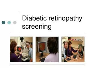
GOLD STANDARD FOR RETINAL SCREENING Retinal photography is the gold standard for screening diabetic retinopathy Seven 30 - degree field of stereoscopic photographs taken by a trained technician Photographs can be taken by a mobile unit with a camera and later assessed by a trained reader Suited to serve even rural communities
SPECIFICITY AND SENSITIVITY OF OPHTHALMOSCOPY AND PHOTOGRAPHY Ophthalmoscopy Photography (%) (%) Sensitivity 65.7 87.3 Specificity 93.8 84.8 Owens et al, Diabetic Medicine, 1998
WHO CAN DO SCREENING ? • General practitioner • Optometrists • Clinicians in a hospital - based diabetes centre • Ophthalmologists • Diabetologists • Retinal photography services • Combination of all these
ERROR RATES FOR DIAGNOSING DIABETIC EYE DISEASE - OPHTHALMOSCOPY Overall Serious errors (%) errors (%) Internist 74 70 Senior medical resident 69 52 Diabetologist 66 50 Ophthalmologist 48 11 Retinal specialist 13 0
NATURAL HISTORY OF NEPHROPATHY IN TYPE 1 DIABETES Stage of hyper- filtration Normo albumi- nuria Micro albumi- nuria Macro albumi- nuria Azotemia (Renal failure) End stage Renal disease 15 - 20 yrs 4 - 5 yrs 1 yrs
PREVALENCE OF DIABETIC NEPHROPATHY Diabetic Nephropathy • Develops in 35 - 45% of Type 1 diabetic patients • 20 - 30% of Type 2 diabetic patients • Leading cause of ESRD in United States
PREVALENCE OF DIABETIC NEPHROPATHY IN DIFFERENT ETHNIC GROUPS 19 million Indians with diabetes 5 - 60% of type 2 diabetes depending on ethnic origin Caucasians - 5 - 10% African Americans - 10 - 20% Pima Indians - 60% Asian Indians - 10% Even with 10%, 1.7 million Indian diabetics will have Nephropathy
SCREENING FOR MICROALBUMINURIA Routine urinalysis for protein Overt nephropathy Quantitative protein begin treatment + For protein - For protein Condition that may invalidate urine albumin excretion Yes Wait until resolved No No Repeat in 1 year Test for microalbumin > 30 mg/24h Yes Repeat microalbumin test twice within 3 months period 2 of 3 tests > 30 mg/24h ? Yes Microalbuminuria, begin treatment
SPECIMEN COLLECTION • Collect freshly voided urine in a clean, dry container • Preservatives should be avoided • Samples which cannot be tested within 3 days of collection should be refrigerated • Samples should not be frozen • The test should be free from significant interference from glucosuria, pH, ketonuria or bacterial contamination
SCREENING FOR MICROALBUMINURIA Three methods Albumin to creatinine ratio in random spot collection 24 - h urine collection with creatinine Timed collection (4-h or overnight)
DEFINITION OF MICROALBUMINURIA Stage 24h Timed Spot collection collection collection Normoalbuminuria < 30 mg/24h <20g/min <30g/mg creat Microalbuminuria 30-300 mg/24h 20-200g/min 30-300g/mg creat Clinical albuminuria >300 mg/24h >200g/min >300g/mg creat ADA, Diabetes Care, 1998
ADVANTAGES AND DISADVANTAGES METHODS OF MICROALBUMINURIA ANALYSIS Random spot collection Easy to perform Generally provides accurate information First void or morning collection Preferred due to diurnal variation in albumin excretion Gold standard Notoriously labour and time intensive Patients co-operation difficult Timed collection
SPECIFICITY AND SENSITIVITY FOR MICROALBUMINURIA Timed urine collection - gold standard Sensitivity Specificity (%) (%) Random spot specimen 89 85 First morning void 70 93 Schwab et al, Diabetes Care, 1992
SHORTENED TIMED CLEARANCES SUGGESTIONS ….. 3 -hour collections Brodows et al, Diabetes Care, 1981 4 -hour collections Steno study group, Lancet, 1982 1 -hour timed collections Sochett et al, J.Pediatr,1988 Overnight collections Viberti et al , Lancet, 1982
ASSAYS FOR MICROALBUMINURIA Qualitative Dipstick method Quantitative Radioimmuno assay Immunoturbidometric assay Enzyme linked Immunosorbant assay
MICRAL STRIPS Micral strip screening tests offer a cost-effective method of screening Dip sticks show acceptable sensitivity (95%) and specificity (93%) All positive tests should be confirmed by more specific methods
FALSE POSITIVES FOR ALBUMINURIA Hyperfiltration (Newly diagnosed diabetes) Exercise Marked hypertension Congestive Heart Failure Urinary Tract Infection Acute febrile illness
CONCLUSIONS Screening for retinopathy Sensitive, specific and safe screening tests are available for retinopathy Retinal photography is the gold standard, which can be modified from seven to four field Training is necessary to grade retinal photographs Newer technologies including digital imaging may reduce the cost of screening
PRIORITIES For preventing blindness due to diabetes • Screening • Diagnosis • Treatment • Counseling • Education For all diabetic patients
CONCLUSIONS Screening for nephropathy Screening tests for microalbuminuria are safe, simple at the same time specific and sensitive Timed urine collection is the gold standard. However spot urine testing has also proved to be equally sensitive Micral dip sticks are cost effective Microalbuminuria provides information not only about nephropathy,but also generalized vascular disease (endothelial dysfunction)
PRIORITIES For preventing nephropathy due to diabetes • Annual screening of Microalbuminuria • Glycemic control • Treatment modalities to slow down the rate of progression of nephropathy in all diabetic patients